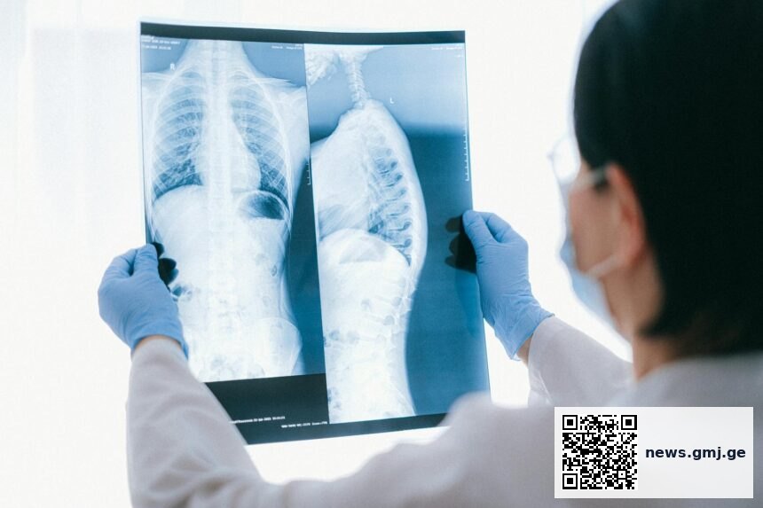
A new intelligence/" class="gmj-dict-autolink" title="Dictionary: Artificial Intelligence">artificial intelligence system can identify early bone loss from routine chest X-rays, potentially transforming osteoporosis screening for millions of patients who fall outside current guidelines. The technology addresses a critical gap where younger adults, men, and normal-weight individuals often remain undiagnosed until after a fracture occurs.
Current osteoporosis screening gaps by demographic group
Percentage of at-risk adults excluded from routine screening guidelines
Source: National Osteoporosis Foundation, 2024 | Georgian Medical Journal News
Silent Disease Affects Diverse Populations
Osteoporosis develops gradually as bones lose density and strength, often progressing undetected for years before the first fracture reveals the underlying condition. Current screening guidelines from the US Preventive Services Task Force primarily target women aged 65 and older, plus younger postmenopausal women with risk factors.

This approach leaves significant populations without routine screening access, including most men under 70, younger women, and individuals who don’t fit traditional risk profiles. The National Osteoporosis Foundation estimates that approximately 10.2 million Americans have osteoporosis, while another 43.4 million have low bone density that puts them at increased fracture risk.
Healthcare systems worldwide face similar challenges in identifying at-risk patients before fractures occur. The development represents a potential shift toward opportunistic screening that could identify bone loss during routine medical imaging. Related advances in clinical diagnostic tools continue to expand early detection capabilities across various conditions.
AI Technology Analyzes Existing Imaging
The artificial intelligence system analyzes chest X-rays that patients receive for other medical reasons, such as pneumonia screening or pre-surgical evaluations. By examining bone density patterns visible in these routine images, the technology can flag patients who may benefit from formal bone density testing.
This approach leverages imaging that healthcare systems already perform routinely, potentially eliminating the need for additional appointments or specialized tests for initial screening. The Centers for Disease Control and Prevention notes that many fractures occur in people who haven’t been diagnosed with osteoporosis, highlighting the importance of broader screening approaches.
Machine learning algorithms trained on thousands of imaging studies can detect subtle changes in bone architecture that might not be apparent to human observers reviewing chest X-rays for other purposes. This represents part of broader trends in artificial intelligence applications for medical diagnosis and screening.
Expanding Access Beyond Current Guidelines
Traditional bone density screening using dual-energy X-ray absorptiometry (DEXA) scans requires specialized equipment and dedicated appointments, creating barriers for some patients. The World Health Organization estimates that osteoporotic fractures will continue increasing globally as populations age, making early detection increasingly important.
Men represent a particularly underserved group in osteoporosis screening, despite accounting for approximately 20% of cases. The condition often goes unrecognized in male patients until severe complications develop. Younger adults and those with normal body weight also fall outside typical screening protocols, even though genetic factors, medications, or underlying conditions may increase their fracture risk.
Integration of AI-based screening into routine chest imaging workflows could identify these overlooked populations without requiring changes to current clinical protocols. This aligns with growing emphasis on preventive healthcare approaches that catch diseases before symptomatic stages develop.
The AI system can detect bone density patterns in routine chest X-rays that correlate with osteoporosis risk, potentially identifying millions of unscreened patients during regular healthcare encounters.
— Medical imaging researchers (Medical Xpress, 2026)
Key takeaways
- AI technology can analyze routine chest X-rays to identify early bone loss before fractures occur
- Current screening guidelines miss significant populations including younger adults, men, and normal-weight individuals
- The approach leverages existing imaging workflows without requiring additional appointments or specialized tests
Frequently asked questions
How accurate is AI screening compared to standard bone density tests?
While specific accuracy data wasn’t provided in the source report, AI screening serves as an initial flag to identify patients who may benefit from formal DEXA scan evaluation. It’s designed to supplement, not replace, standard diagnostic methods.
Who would benefit most from this AI screening approach?
Patients currently excluded from routine osteoporosis screening guidelines would benefit most, including men under 70, women under 50-65, and individuals without traditional risk factors who receive chest X-rays for other medical reasons.
When might this technology become available in clinical practice?
The source article didn’t specify implementation timelines, but medical AI tools typically require regulatory approval and clinical validation studies before becoming widely available in healthcare systems.
As healthcare systems seek more efficient ways to identify disease risk in diverse populations, AI-powered analysis of routine imaging represents a promising avenue for expanding preventive care access. The technology’s ability to work with existing clinical workflows could help address current gaps in osteoporosis screening while supporting broader goals of early disease detection and fracture prevention.
Source: AI repurposes routine chest X-rays to catch silent bone loss before fracture
Was this article helpful?
Disclaimer. This article is health journalism intended for general information and education. It is not medical advice and is not a substitute for professional diagnosis or treatment. Always consult a qualified healthcare provider about your individual circumstances. Full disclaimer →
Related Coverage


Editorial standards. This article was produced under the GMJ News editorial process, with oversight by the GMJ Editorial Board. Our editorial process. Spotted an error? Contact the editorial team.