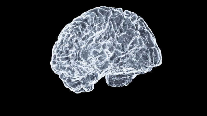
A significant clinical gap exists in current brain tumor detection: approximately one in four patients with detectable tumors receive normal results on standard MRI. Stanford researchers have now demonstrated that enhanced contrast MRI can bridge this diagnostic gap, identifying tumors as small as 2 millimeters compared to the conventional 5-millimeter threshold.
For patients with suspected brain tumors or concerning neurological symptoms, these findings suggest that negative standard MRI results may not conclusively exclude a tumor diagnosis. Enhanced contrast MRI may warrant consideration, particularly when clinical suspicion remains high. The 23% detection rate in patients with normal conventional scans underscores the importance of diagnostic imaging evolution.
Early tumor detection has direct bearing on treatment outcomes and survival. The potential to improve glioblastoma survival rates from 6.8% to 23.4% through earlier intervention represents substantial clinical value. As multicenter trials begin in 2027, enhanced contrast MRI may become standard protocol in neuro-oncology departments, fundamentally improving diagnostic accuracy and patient care trajectories.
Read the full article on GMJ Newsroom.

Was this article helpful?