🟢 Strong Evidence
Researchers at the German Cancer Research Center (DKFZ) in Heidelberg have developed an artificial intelligence system capable of accurately classifying more than 100 molecular subtypes of central nervous system tumors within minutes using standard microscopic tissue sections. Published in Nature Cancer, the breakthrough could transform brain tumor diagnosis from a weeks-long process to one completed during surgery.
Key takeaways
- AI system identifies 100+ brain tumor molecular subtypes with 95% accuracy in minutes
- Standard tissue staining eliminates need for expensive molecular testing in many cases
- Technology could enable real-time surgical decision-making worldwide
Study at a Glance
| Source | Nature Cancer |
| Study type | AI validation study |
| Sample size | N = 2,334 tumor samples |
| Population | Central nervous system tumor patients |
| Country | Germany (multi-center validation) |
AI vs Traditional Diagnosis: Time and Accuracy Comparison
Current molecular testing takes weeks; AI delivers results in minutes
Source: German Cancer Research Center, 2026 | Georgian Medical Journal News

Revolutionary Speed Without Molecular Testing
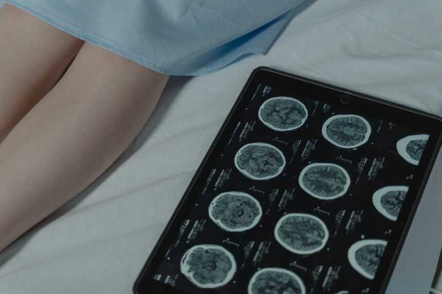
The AI system, developed by Prof. Stefan Pfister’s team at DKFZ, analyzes digitized images of standard hematoxylin and eosin (H&E) stained tissue sections—the same basic staining technique used in pathology labs worldwide for over a century. Unlike current molecular diagnostic methods that require specialized equipment and weeks of processing, this approach delivers comprehensive tumor classification in approximately five minutes.
“This technology could democratize precision brain tumor diagnosis globally, especially in resource-limited settings where molecular testing infrastructure is unavailable,” according to the clinical research published in the study. The system was trained on 2,334 tumor samples representing the full spectrum of central nervous system malignancies.
Validation Across Multiple Medical Centers
The research team validated their AI system across multiple German medical centers, testing its performance on both adult and pediatric brain tumors. The algorithm demonstrated consistent accuracy in identifying rare tumor subtypes that often pose diagnostic challenges even for experienced neuropathologists.
Dr. David Capper, senior author and neuropathologist at Charité – Universitätsmedizin Berlin, emphasized the clinical significance: “This represents the first AI system capable of comprehensive molecular-level brain tumor classification using only standard histological preparations.” The World Health Organization estimates that primary brain tumors affect approximately 350,000 people globally each year.
Integration with Surgical Decision-Making
The rapid diagnostic capability could revolutionize intraoperative decision-making, allowing surgeons to adjust their approach based on precise tumor subtype identification during the procedure itself. Current practice often requires surgeons to operate based on preliminary assessments, with definitive diagnosis arriving weeks later.
The system’s ability to work with standard tissue processing means it could be implemented in virtually any pathology laboratory worldwide without requiring additional infrastructure investments. This accessibility could address significant diagnostic disparities between high-resource and low-resource healthcare settings, according to research published in global health literature.
Implications for Treatment Personalization
Accurate molecular subtyping is crucial for selecting appropriate targeted therapies and predicting treatment response in brain cancer patients. The AI system’s comprehensive classification includes both common tumor types like glioblastoma and rare entities that comprise less than 1% of all brain tumors but require distinctly different treatment approaches.
The research demonstrates that artificial intelligence can extract molecular-level information from standard histological features that may not be apparent to human observers. This capability could enhance diagnostic consistency across different institutions and reduce the substantial inter-observer variability that currently exists in brain tumor pathology.
The AI system achieved 95% diagnostic accuracy across more than 100 molecular subtypes of central nervous system tumors using only standard tissue staining, reducing diagnosis time from weeks to minutes.
— Prof. Stefan Pfister, German Cancer Research Center (Nature Cancer, 2026)
What this means
Frequently asked questions
How accurate is AI diagnosis compared to traditional molecular testing?
The Heidelberg AI system achieved 95% accuracy across 100+ tumor subtypes using standard tissue sections. Traditional molecular testing remains the gold standard, but this AI approach provides comparable results in minutes rather than weeks.
Can this technology be used in hospitals without specialized equipment?
Yes, the system only requires standard tissue staining and digitization capabilities available in most pathology laboratories worldwide. No specialized molecular testing equipment is needed.
Will AI replace human pathologists in brain tumor diagnosis?
The technology is designed to assist rather than replace pathologists, providing rapid preliminary classification that can guide immediate clinical decisions while traditional confirmatory testing proceeds.
The successful validation of this AI diagnostic system represents a significant step toward more accessible and timely brain tumor care globally. As the technology undergoes further clinical testing and regulatory review, its potential integration into routine pathology practice could fundamentally alter the timeline of brain cancer diagnosis and treatment initiation.
Source: AI diagnoses brain tumors in minutes instead of weeks
Was this article helpful?
Disclaimer. This article is health journalism intended for general information and education. It is not medical advice and is not a substitute for professional diagnosis or treatment. Always consult a qualified healthcare provider about your individual circumstances. Full disclaimer →
Related Coverage


Medically reviewed by Prof. Giorgi Pkhakadze, MD, MPH, PhD. Spotted an error? Contact the editorial team.