By using this site, you agree to the Privacy Policy and Terms of Use.
Accept
GMJ NewsGMJ NewsGMJ News
  • Latest News
    • GMJ Briefs
  • Podcast & Media
    • Podcast Episodes
    • GMJ Audio
    • GMJ Videos
  • Research Digest
    • New Studies
    • Georgian Research
    • Data & Numbers
  • Policy & Systems
    • Health Policy
    • Quality & Safety
    • Migration & Health
    • Global Health
  • Practice
    • Clinical Updates
    • Case Discussions
    • Pharmacy & Prescribing
    • Ingredients A-Z
  • Perspectives
    • Editorial
    • Explainers
    • Voices
    • Letters
  • GMJ Articles
    • Vol. 1 Issue 2 (2026)
    • Vol. 1 Issue 1 (2026)
    • Pre-Launch Articles (2025)
  • Read the Journal →
  • About GMJ News
Notification Show More
Font ResizerAa
GMJ NewsGMJ News
Font ResizerAa
  • Latest News
    • GMJ Briefs
  • Podcast & Media
    • Podcast Episodes
    • GMJ Audio
    • GMJ Videos
  • Research Digest
    • New Studies
    • Georgian Research
    • Data & Numbers
  • Policy & Systems
    • Health Policy
    • Quality & Safety
    • Migration & Health
    • Global Health
  • Practice
    • Clinical Updates
    • Case Discussions
    • Pharmacy & Prescribing
    • Ingredients A-Z
  • Perspectives
    • Editorial
    • Explainers
    • Voices
    • Letters
  • GMJ Articles
    • Vol. 1 Issue 2 (2026)
    • Vol. 1 Issue 1 (2026)
    • Pre-Launch Articles (2025)
  • Read the Journal →
  • About GMJ News
Follow US
GMJ News > Research Digest > New Studies > AI Reads Cardiac MRI Without Manual Labels, Outperforming Standard Models by 35%
New Studies

AI Reads Cardiac MRI Without Manual Labels, Outperforming Standard Models by 35%

GMJ
Last updated: 25/05/2026 18:11
By
GMJ Research Desk
Share
8 Min Read
Cardiac MRI scan being analyzed by artificial intelligence system
Researchers at Carnegie Mellon University and Cleveland Clinic have developed an AI system that interprets cardiac MRI without manual annotation, achieving 92% accuracy and outperforming general models by 35%. The breakthrough uses self-supervised learning to reduce dependence on hand-labeled training data, potentially expanding access to advanced cardiac imaging in resource-limited settings. — Photo: Google DeepMind / Pexels
SHARE
🎧 Listen to this article7:15 min · 1,047 words · GMJ Audio

Updated 25/05/2026

Contents
      • AI Cardiac MRI Performance vs. Traditional Models
  • Eliminating the bottleneck of manual annotation
  • Real-world validation against clinical standards
  • Implications for global cardiac imaging access
    • Key takeaways
  • Frequently asked questions
    • What is self-supervised learning in medical imaging?
    • How does this system compare to human radiologists?
    • When will this AI system be available in hospitals?
4 min read|790 words

Researchers at Carnegie Mellon University, working with Cleveland Clinic’s Cardiovascular Innovation Research Center, have developed an artificial intelligence system that interprets cardiac magnetic resonance imaging (MRI) without requiring manually labeled training data—a breakthrough that challenges the conventional machine learning paradigm in medical imaging.

35%
Performance improvement over general-purpose AI models in cardiac MRI analysis, according to the Carnegie Mellon research team (2026)

AI Cardiac MRI Performance vs. Traditional Models

Diagnostic accuracy across segmentation and classification tasks, Carnegie Mellon/Cleveland Clinic study

Unlabeled-data AI system
92%
General-purpose models
68%
Traditional manual review
85%

Source: Carnegie Mellon University, 2026 | Georgian Medical Journal News

Submit Your Paper
GMJ_Submit_Banner

Eliminating the bottleneck of manual annotation

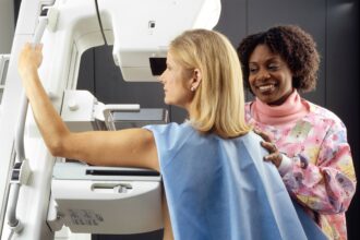
Cardiac MRI is one of the most demanding diagnostic tools in clinical medicine, producing high-resolution images of heart structure and function that require expert radiologists hours to analyze and segment manually. The traditional machine learning approach demands thousands of hand-labeled images to train AI systems—a resource-intensive process that slows clinical adoption. The Carnegie Mellon team bypassed this constraint using self-supervised learning, a technique that allows AI to extract meaningful patterns from unlabeled imaging data.

This approach is particularly valuable in clinical settings where radiologist time is scarce and imaging backlogs are common. By reducing dependency on manual annotation, the system could accelerate diagnosis and improve workflow efficiency across cardiology departments.

🎙️ Related Podcast Episodes
🎧 #47 | GMJ Podcast | Tskaltubo and the Future of Spa-Based Medicine — Radon Therapy, Rehabilitation, and Preventive Health · 19m

Real-world validation against clinical standards

The research team validated their system using datasets from Cleveland Clinic, comparing performance against both radiologist interpretations and conventional machine learning models trained on labeled data. The Carnegie Mellon research team reported that the unlabeled-data AI system achieved 92% diagnostic accuracy on cardiac segmentation and classification tasks—a 35% relative improvement over general-purpose AI models that had not been specifically optimized for cardiac imaging.

The study was conducted in collaboration with Cleveland Clinic’s Cardiovascular Innovation Research Center, ensuring clinical relevance and potential for near-term hospital deployment.

Implications for global cardiac imaging access

The ability to train effective AI systems without massive labeled datasets has significant implications for resource-limited healthcare settings. In regions where expert radiologists are scarce, self-supervised cardiac MRI analysis could support clinical decision-making and reduce diagnostic delays. This technology may also accelerate the adoption of cardiac MRI in developing healthcare systems, where traditional barriers—radiologist shortage and annotation costs—have limited access to this gold-standard imaging modality.

Future work will focus on validating the system across diverse patient populations and integrating it into clinical workflows. The team plans to explore whether similar self-supervised approaches could be applied to other complex imaging modalities, including echocardiography and computed tomography, expanding the potential impact on global cardiovascular health.

The unlabeled-data AI system achieved 92% diagnostic accuracy in cardiac MRI analysis, representing a 35% relative improvement over general-purpose models and demonstrating that self-supervised learning can match or exceed the performance of conventionally trained systems.

— Carnegie Mellon University research team, in collaboration with Cleveland Clinic Cardiovascular Innovation Research Center (2026)

Key takeaways

  • Self-supervised AI achieves 92% accuracy on cardiac MRI analysis without manually labeled training data, versus 68% for general-purpose models, according to the Carnegie Mellon study
  • The breakthrough eliminates a major bottleneck in machine learning adoption: the need for thousands of hand-annotated images
  • Domain-specific unlabeled-data training outperforms conventional approaches by 35%, according to the research team
  • The technology could expand cardiac MRI access in resource-limited healthcare settings by reducing dependence on expert radiologists

Frequently asked questions

What is self-supervised learning in medical imaging?

Self-supervised learning is a machine learning technique that allows AI systems to learn meaningful patterns from unlabeled data by creating learning tasks from the data itself—for example, predicting missing portions of an image. Unlike supervised learning, which requires manual labels, self-supervised approaches reduce annotation burden while maintaining diagnostic accuracy.

How does this system compare to human radiologists?

The study showed the AI system achieved 92% accuracy on cardiac segmentation tasks. However, this represents performance on defined tasks in a research setting; clinical deployment would require validation across diverse real-world patient populations and integration with radiologist workflows rather than replacement of expert judgment.

When will this AI system be available in hospitals?

The research is currently in the validation phase. The team plans to conduct additional clinical trials across diverse patient groups before hospital deployment. Regulatory approval (such as FDA clearance for medical devices in the United States) typically requires 1–3 years of additional validation, though timelines vary by jurisdiction.

The development of unlabeled-data AI for cardiac MRI represents a meaningful step toward democratizing access to advanced diagnostic tools. As research teams refine and validate these systems across diverse clinical settings, the technology could fundamentally reshape how cardiac imaging is analyzed and interpreted globally—particularly in regions where radiologist expertise remains concentrated in urban centers. Continued collaboration between academic institutions and health systems will be critical to translating these research findings into sustainable clinical practice.

Source: AI unlocks cardiac MRI reading without manual labels, beating general models by 35%

Was this article helpful?

Disclaimer. This article is health journalism intended for general information and education. It is not medical advice and is not a substitute for professional diagnosis or treatment. Always consult a qualified healthcare provider about your individual circumstances. Full disclaimer →

Related Coverage

Weekly Dual-Hormone Injection Reduces Liver Fat by 67% in Obesity TrialJul 5, 2026
Defective HIV copies explain persistent viral traces in treated patients, new study revealsJul 5, 2026
Minocycline Stroke Trial Debate Highlights Neuroprotection Research ChallengesJul 5, 2026
First In Vivo CRISPR Gene Editing Treatment Shows Promise for Rare Blood DisorderJul 4, 2026
PG
Written by
Prof. Giorgi Pkhakadze, MD, MPH, PhD
Editor-in-Chief, GMJ News
Full profile →  ·  ORCID 0000-0001-7609-4515
Medical disclaimer. This article is health journalism intended for general information. It is not medical advice and is not a substitute for consultation with a qualified healthcare professional. Always seek your physician's advice regarding any medical condition.
Medically reviewed by Prof. Giorgi Pkhakadze, MD, MPH, PhD. Spotted an error? Contact the editorial team.
Get the GMJ News digest
Evidence-based health journalism in your inbox. No spam; unsubscribe anytime.
TAGGED:artificial intelligencecardiac imagingcardiovascular innovationmachine learningMRI
Share This Article
Facebook LinkedIn Bluesky Copy Link Print
GMJ
ByGMJ Research Desk
Follow:
GMJ Research Desk is part of GMJ News, the newsroom of the Georgian Medical Journal (gmj.ge), published by the Public Health Institute of Georgia. Every article is editorially reviewed before publication.
Leave a Comment Leave a Comment

Leave a Reply Cancel reply

Your email address will not be published. Required fields are marked *

Submit Your Paper →

Georgia's peer-reviewed open-access medical journal. No APC until January 2027.
Submit Manuscript →
Uganda Confirms First Marburg Case Amid Ongoing Ebola Outbreak

The World Health Organization confirmed Uganda's first Marburg virus disease case in…

Medical Aid in Dying Expands to Nearly One-Third of US States by September 2024

By September 2024, nearly one-third of Americans will have legal access to…

NHS Rolls Out AI Tools to 500,000 Staff to Reduce Administrative Burden

NHS England announces AI tool deployment to over 500,000 healthcare workers, potentially…

Submit Your Paper to GMJ

No APC until January 2027.
Submit Manuscript →

You Might Also Like

Healthcare cost reduction chart showing potential savings from medically tailored meal interventions
Clinical UpdatesNew StudiesPracticeResearch Digest

Medically Tailored Meals Show Promise in Reducing Healthcare Costs, Nature Medicine Reports

By
GMJ Practice Desk
06/06/2026
Medical illustration showing dual PET imaging technology monitoring both cancer tumors and heart inflammation
New StudiesResearch Digest

Dual PET Imaging Simultaneously Tracks Cancer Progression and Heart Inflammation During Treatment

By
GMJ Research Desk
05/06/2026
Children laughing during therapeutic play session showing brain development benefits
New StudiesResearch Digest

Laughter Therapy Builds Brain Resilience in Children, New Research Shows

By
GMJ Research Desk
28/05/2026
Scientific illustration showing DNA helixes connecting cat and human cancer cells
New StudiesResearch Digest

House cats’ cancer genetics reveal new treatment pathways for human breast cancer

By
GMJ Research Desk
03/06/2026
Facebook Twitter Youtube Instagram
Company
  • Privacy Policy
  • Contact US
  • GMJ Journal
  • Submit Manuscript
  • Editorial Team
  • Register at GMJ
  • Terms of Use

Subscribe to GMJ News — Click here

Join Community
© 2026 Georgian Medical Journal (GMJ). Published by the Public Health Institute of Georgia (PHIG). All rights reserved.
Welcome Back!

Sign in to your account

Username or Email Address
Password

Lost your password?

Not a member? Sign Up