🟡 Preliminary Evidence
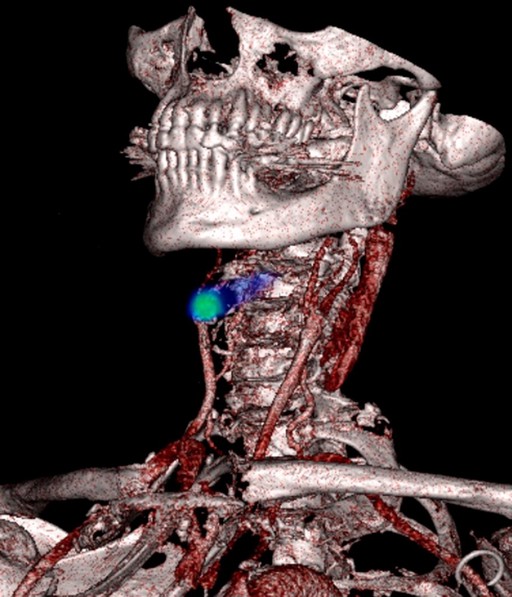
A novel positron emission tomography (PET) radiotracer can simultaneously detect deep vein thrombosis in the legs and identify whether blood clots have migrated to the lungs, according to research presented at the Society of Nuclear Medicine and Molecular Imaging (SNMMI) 2026 Annual Meeting. The breakthrough imaging approach enables whole-body visualization of blood clots in a single scan, potentially revolutionizing diagnosis of venous thromboembolism. The collection of images demonstrating blood clot visualization has been selected as the 2026 SNMMI Henry N. Wagner, Jr., Image of the Year.
Key takeaways
- New PET radiotracer accurately detects deep vein thrombosis and pulmonary embolism in one scan
- Whole-body imaging approach may reduce diagnostic time and improve patient outcomes
- Research findings recognized with prestigious 2026 SNMMI Image of the Year award
Venous Thromboembolism: A Major Health Challenge
Annual incidence and mortality impact globally
Source: European Centre for Disease Prevention and Control, 2022 | Georgian Medical Journal News

Revolutionary Imaging Technology Shows Promise
The novel PET radiotracer represents a significant advancement in thrombosis detection, addressing current limitations in diagnostic imaging. Traditional approaches typically require separate imaging studies for suspected deep vein thrombosis and pulmonary embolism, potentially delaying treatment decisions. Research presented at the SNMMI meeting demonstrates the tracer’s ability to provide comprehensive visualization of the venous system in a single procedure.
Current diagnostic protocols often involve duplex ultrasound for lower extremity evaluation and computed tomography pulmonary angiography (CTPA) for lung assessment. The Centers for Disease Control and Prevention estimates that venous thromboembolism affects up to 900,000 Americans annually, with pulmonary embolism causing approximately 100,000 to 180,000 deaths per year.
Clinical Impact and Diagnostic Efficiency
The whole-body imaging capability addresses a critical clinical need for patients presenting with symptoms that could indicate either deep vein thrombosis or pulmonary embolism. Emergency department physicians frequently encounter patients with chest pain, shortness of breath, or leg swelling where the source of potential thrombosis remains unclear. A comprehensive single-scan approach could streamline the diagnostic process and reduce radiation exposure compared to multiple imaging studies.
According to the World Health Organization, cardiovascular diseases remain the leading cause of death globally, with thrombotic events contributing significantly to this burden. The novel tracer technology may enable earlier detection and treatment initiation, potentially improving patient outcomes. For more insights into cutting-edge diagnostic technologies, see our New Studies section.
Recognition and Future Clinical Applications
The selection of this research as the 2026 SNMMI Image of the Year underscores its potential clinical significance. The Henry N. Wagner, Jr., Image of the Year award recognizes outstanding contributions to nuclear medicine imaging that demonstrate exceptional scientific merit and clinical relevance.
The imaging approach may prove particularly valuable in complex clinical scenarios where patients present with multiple risk factors for thrombotic disease. This includes hospitalized patients, those with cancer, individuals with inherited thrombophilia, or patients receiving hormone therapy. The published literature on advanced thrombosis imaging continues to evolve, with researchers exploring various molecular targets for improved detection.
The novel PET radiotracer enables simultaneous detection of deep vein thrombosis and pulmonary embolism in a single whole-body scan, potentially transforming diagnostic approaches for venous thromboembolism.
— Research presented at Society of Nuclear Medicine and Molecular Imaging 2026 Annual Meeting
What this means
Frequently asked questions
How does this new PET tracer differ from current imaging methods?
The novel tracer provides whole-body visualization of blood clots in a single scan, unlike current approaches that typically require separate ultrasound for legs and CT scanning for lungs. This comprehensive approach may reduce diagnostic time and improve detection accuracy.
What are the current challenges in diagnosing blood clots?
Current diagnostic protocols often involve multiple imaging studies, which can delay treatment decisions and increase patient radiation exposure. Traditional methods may miss clots in certain locations or require clinical judgment about which body regions to image.
When might this technology become available for clinical use?
While the research shows promising results, the technology requires further clinical validation and regulatory approval before routine clinical implementation. The timeline for availability will depend on additional safety and efficacy studies.
The recognition of this imaging advance highlights the ongoing innovation in nuclear medicine and molecular imaging approaches to thrombotic disease. As research continues to validate the clinical utility of novel PET tracers, the potential for improved patient care through more efficient and comprehensive diagnostic approaches becomes increasingly apparent. This technological advancement represents an important step toward personalized medicine approaches for venous thromboembolism management.
Source: New PET tracer identifies DVT in legs and lungs
Was this article helpful?
Disclaimer. This article is health journalism intended for general information and education. It is not medical advice and is not a substitute for professional diagnosis or treatment. Always consult a qualified healthcare provider about your individual circumstances. Full disclaimer →
Related Coverage


Medically reviewed by Prof. Giorgi Pkhakadze, MD, MPH, PhD. Spotted an error? Contact the editorial team.