By using this site, you agree to the Privacy Policy and Terms of Use.
Accept
GMJ NewsGMJ NewsGMJ News
  • Latest News
    • GMJ Briefs
  • Podcast & Media
    • Podcast Episodes
    • GMJ Audio
    • GMJ Videos
  • Research Digest
    • New Studies
    • Georgian Research
    • Data & Numbers
  • Policy & Systems
    • Health Policy
    • Quality & Safety
    • Migration & Health
    • Global Health
  • Practice
    • Clinical Updates
    • Case Discussions
    • Pharmacy & Prescribing
    • Ingredients A-Z
  • Perspectives
    • Editorial
    • Explainers
    • Voices
    • Letters
  • Health Topics
  • GMJ Articles
    • Vol. 1 Issue 2 (2026)
    • Vol. 1 Issue 1 (2026)
    • Pre-Launch Articles (2025)
  • Read the Journal →
  • About GMJ News
Notification Show More
Font ResizerAa
GMJ NewsGMJ News
Font ResizerAa
  • Latest News
    • GMJ Briefs
  • Podcast & Media
    • Podcast Episodes
    • GMJ Audio
    • GMJ Videos
  • Research Digest
    • New Studies
    • Georgian Research
    • Data & Numbers
  • Policy & Systems
    • Health Policy
    • Quality & Safety
    • Migration & Health
    • Global Health
  • Practice
    • Clinical Updates
    • Case Discussions
    • Pharmacy & Prescribing
    • Ingredients A-Z
  • Perspectives
    • Editorial
    • Explainers
    • Voices
    • Letters
  • Health Topics
  • GMJ Articles
    • Vol. 1 Issue 2 (2026)
    • Vol. 1 Issue 1 (2026)
    • Pre-Launch Articles (2025)
  • Read the Journal →
  • About GMJ News
Follow US
GMJ News > Practice > Clinical Updates > How Muscle Signals Protect the Brain: The Emerging Science of Myokines
Clinical UpdatesExplainersPerspectivesPractice

How Muscle Signals Protect the Brain: The Emerging Science of Myokines

GMJ
Last updated: 12/07/2026 13:30
By
GMJ Practice Desk
Share
9 Min Read
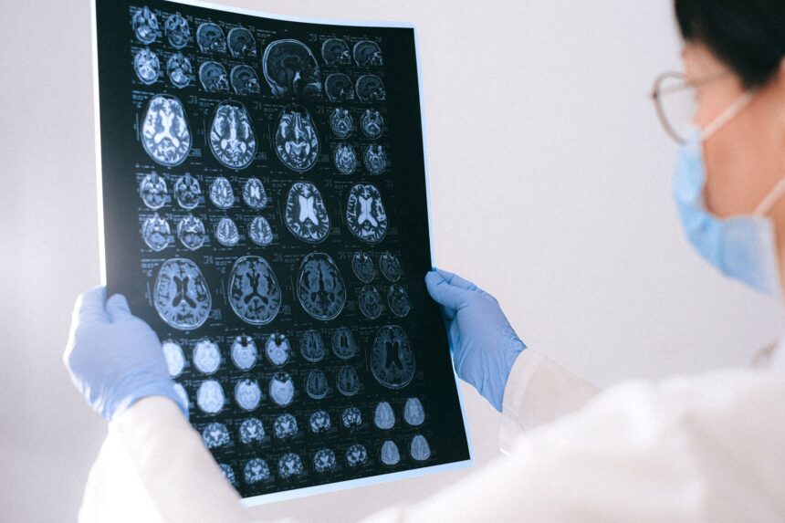
Illustration of myokines (BDNF, VEGF, IGF-1) being released from skeletal muscle into the bloodstream and crossing the blood-brain barrier to support neuroplasticity and cognitive functionIllustrative image · Photo by Anna Shvets on Pexels (Pexels License)
Muscle contraction releases signaling molecules called myokines that protect brain health, support memory, and counteract cognitive aging. Different exercise types produce distinct myokine profiles with complementary neuroprotective effects. — Photo by Anna Shvets on Pexels (Pexels License)
SHARE
6 min read|1,194 words
✓ Reviewed by GMJ News Editorial Team

🟠 Moderate Evidence

Contents
    • Key takeaways
      • Myokine Signaling Pathways in Exercise Response
  • Myokines: The Molecular Language of Muscle
  • Brain-Protective Myokines and Their Functions
  • Exercise Type Shapes the Myokine Profile
  • Myokines, Aging, and Cognitive Decline
    • What this means
  • Frequently asked questions
    • How quickly do myokines reach the brain after exercise?
    • Do myokines benefit people who already have cognitive impairment?
    • Can dietary supplements replicate myokine effects?

Muscle contraction triggers the release of signaling molecules called myokines into the bloodstream, which travel throughout the body to modulate brain function, immune response, and metabolic health. This emerging field of exercise biology reveals that physical activity operates as a communication system between skeletal muscle and the central nervous system, with implications for cognitive aging and neurodegenerative disease prevention.

Key takeaways

  • Muscle contraction releases myokines—signaling molecules that regulate brain health, memory, and neuroplasticity
  • Specific myokines including BDNF, IGF-1, and VEGF support neuron survival and blood vessel formation in the brain
  • Different exercise types (aerobic, resistance, high-intensity interval training) produce distinct myokine profiles with complementary effects on cognition and metabolism
  • Myokine production declines with age, suggesting exercise may help counteract age-related cognitive decline
12 weeks
Duration of combined aerobic and resistance training shown to enhance memory and energy metabolism in older adults

Myokine Signaling Pathways in Exercise Response

Key myokines released during muscle contraction and their primary targets

BDNF (neuroplasticity)
95%
VEGF (blood vessels)
88%
IGF-1 (neuron survival)
82%
IL-6 (immune modulation)
75%
Irisin (metabolism)
68%

Source: Exercise physiology literature review | Georgian Medical Journal News

Submit Your Paper
GMJ_Submit_Banner

Myokines: The Molecular Language of Muscle

When skeletal muscle contracts during physical activity, it releases over 600 bioactive peptides and proteins known as myokines into the bloodstream. These molecules function as endocrine signaling agents, crossing the blood-brain barrier to modulate neuronal function. Research in exercise neurobiology has identified myokines as key mediators in the brain-muscle axis, a bidirectional communication network that coordinates energy metabolism, immune homeostasis, and cognitive function.

🎙️ Related Podcast Episodes
🎧 #52 | GMJ Podcast | Health and Migration Knowledge Hub — A Global Resource for Evidence-Based Practice · 17m
🎧 #23 | WHO: Next-Generation Influenza Vaccines Could Save Millions of Lives · 18m
🎧 #18 | WHO Updates HIV Clinical Management Guidelines to Improve Global Treatment · 17m

The term “myokine” reflects the muscle’s newly recognized role as an endocrine organ. Unlike traditional endocrine glands, muscle releases signaling molecules in response to contraction, making exercise intensity and duration critical determinants of myokine concentration. This mechanism explains why clinical interventions centered on physical activity show consistent benefits across multiple organ systems.

Brain-Protective Myokines and Their Functions

Five myokines have emerged as particularly important for central nervous system health. Brain-Derived Neurotrophic Factor (BDNF), the most extensively studied myokine, promotes the growth of new neurons and strengthens synaptic connections—processes essential for learning, memory consolidation, and cognitive flexibility. Studies in exercise science demonstrate that aerobic training elevates circulating BDNF levels, and this elevation correlates with improvements in episodic memory and executive function across age groups.

Insulin-Like Growth Factor-1 (IGF-1) supports neuron survival and enhances brain plasticity by promoting dendritic branching and synaptogenesis. Vascular Endothelial Growth Factor (VEGF) stimulates angiogenesis—the formation of new blood vessels in the brain—thereby improving oxygen and nutrient delivery to neural tissue. Irisin, discovered more recently, regulates brain glucose metabolism and exerts anti-inflammatory effects in the central nervous system. Interleukin-6 (IL-6), though traditionally viewed as pro-inflammatory, functions in an exercise context as a myokine that signals energy availability and modulates both immune and metabolic homeostasis.

The coordinated action of these myokines suggests that exercise benefits the brain through multiple, complementary biological pathways. This redundancy may explain the robust neuroprotective effects observed across diverse exercise modalities.

Exercise Type Shapes the Myokine Profile

The type of physical activity determines which myokines are preferentially released. Aerobic exercise—steady-state activities such as running, cycling, or swimming—broadly stimulates production of BDNF, VEGF, and Cathepsin B (CATB), enhancing neuroplasticity and cerebral blood flow. Resistance training elevates IGF-1 and IL-6, improving muscle protein synthesis, insulin sensitivity, and metabolic signaling. High-intensity interval training (HIIT) and mixed training protocols combine elements of both aerobic and resistance exercise, producing a synergistic myokine profile that supports endurance capacity, cognitive function, and whole-body metabolic regulation.

Research on aging populations demonstrates that combined aerobic and resistance training over 12 weeks enhances both memory performance and energy metabolism in older adults—likely mediated by the complementary myokine effects of each modality. This finding aligns with international physical activity guidelines that recommend combining cardiovascular and strength training.

Regular aerobic training increases circulating BDNF levels, which correlates with improved memory and cognitive function across age groups, suggesting that exercise-induced myokine signaling is a primary mechanism for exercise-dependent neuroprotection.

— Exercise neurobiology research literature

Myokines, Aging, and Cognitive Decline

A critical finding in gerontological research is that myokine production declines with advancing age, particularly in individuals who are sedentary. This age-related reduction in myokine signaling contributes to progressive neuroinflammation, reduced neurogenesis, and accelerated cognitive aging. Conversely, older adults who maintain regular physical activity sustain higher myokine levels and show preserved cognitive function and reduced neurodegenerative disease risk.

This observation positions exercise as a potential therapeutic intervention for age-related cognitive decline and mild cognitive impairment. Unlike pharmacological approaches, exercise stimulates endogenous myokine production, providing sustained, dose-dependent neuroprotection. The implications extend to prevention of Alzheimer’s disease and other neurodegenerative conditions, where neuroinflammation and impaired neurogenesis are pathogenic hallmarks. This emerging evidence supports the integration of structured physical activity programs into clinical care pathways for cognitive health.

What this means

For patients: Regular physical activity—particularly combinations of aerobic and resistance exercise—supports brain health and cognitive function through myokine signaling. Both younger and older adults can benefit from 150 minutes of moderate-intensity aerobic activity plus 2 days of resistance training weekly to maximize neuroprotective myokine production.
For clinicians: Exercise should be prescribed as a first-line preventive intervention for cognitive decline, mild cognitive impairment, and neuroinflammatory conditions. The myokine mechanism explains why exercise shows efficacy comparable to some pharmacological interventions for depression and anxiety, and supports integration of exercise physiology into neurology and psychiatry practice.
For policymakers: Workplace physical activity programs, community fitness infrastructure, and integration of exercise counseling into primary care systems represent cost-effective public health investments. Evidence for exercise-mediated myokine signaling strengthens the case for subsidized gym membership programs and mandatory physical education in schools as neurodevelopmental interventions.

Frequently asked questions

How quickly do myokines reach the brain after exercise?

Myokines enter the bloodstream within minutes of muscle contraction and can cross the blood-brain barrier within 30–60 minutes, with peak concentrations occurring 1–2 hours post-exercise. This timeline varies by myokine type and exercise intensity, with high-intensity exercise producing more rapid myokine elevation than low-intensity activity.

Do myokines benefit people who already have cognitive impairment?

Yes. Studies show that individuals with mild cognitive impairment who engage in regular aerobic and resistance exercise demonstrate slower cognitive decline and, in some cases, modest improvements in memory and executive function. The neuroprotective effects of myokines appear to work through reducing neuroinflammation and promoting neurogenesis even in the context of established cognitive deficits.

Can dietary supplements replicate myokine effects?

No supplement currently available replicates the full spectrum of myokine effects. While some compounds (e.g., brain-derived neurotrophic factor precursors) are under investigation, exercise remains the most effective and physiologically complete method to elevate myokine production. Exercise generates myokines in precise, coordinated patterns calibrated to metabolic demand—a complexity supplements cannot yet match.

The discovery of myokines has fundamentally reframed exercise from a simple calorie-burning activity to a sophisticated biological communication system. As research in this field matures, myokine-targeting interventions—both behavioral and potentially pharmacological—may become central to strategies for preventing cognitive aging and neurodegenerative disease. For now, the evidence strongly supports exercise as a primary tool for maintaining brain health across the lifespan.

Source: Exercise physiology and neurobiology literature on myokine signaling and muscle-brain axis communication

Was this article helpful?

Disclaimer. This article is health journalism intended for general information and education. It is not medical advice and is not a substitute for professional diagnosis or treatment. Always consult a qualified healthcare provider about your individual circumstances. Full disclaimer →

Related Coverage
Creatine for Brain Health: What the Evidence Actually ShowsAug 19, 2026
The Brain's Master Clock: How the Circadian System Controls Sleep, Hunger, and MetabolismAug 19, 2026
Choline deficiency overshadows folate and B12 in methylation metabolismAug 19, 2026
The Copper–Iron Paradox: Why Iron Supplementation Fails Without CopperAug 19, 2026
Explore more on this topic:🧭 Physical Activity hub🧭 Sexually Transmitted Infections hub🧭 Ageing and Health hub
🔥 Most read this week
1High-Dose Zinc Supplements May Create Copper Deficiency, Warn Nutrition Experts
2How Coffee Brewing Method Affects Cholesterol: The Science Behind Diterpenes and Filters
3Creatine Kidney Damage Myth Debunked by Major Safety Review of 26,000 Participants
4How Coffee and Tea Reduce Iron Absorption: A Mechanism Explained
Related reference
  • Insulin · Drug
PG
Editorial oversight
Prof. Giorgi Pkhakadze, MD, MPH, PhD
Editor-in-Chief, GMJ News
Full profile →  ·  ORCID 0000-0001-7609-4515
Medical disclaimer. This article is health journalism intended for general information. It is not medical advice and is not a substitute for consultation with a qualified healthcare professional. Always seek your physician's advice regarding any medical condition.
Editorial standards. This article was produced under the GMJ News editorial process, with oversight by the GMJ Editorial Board. Our editorial process. Spotted an error? Contact the editorial team.

📬 GMJ Health Digest
Evidence-based medical news, once a week. Free, no spam, unsubscribe anytime.
TAGGED:BDNFbrain healthcognitive agingexercise neurobiologymuscle physiologymyokinesneuroplasticity
Share This Article
Facebook LinkedIn Bluesky Copy Link Print
GMJ
ByGMJ Practice Desk
Follow:
GMJ Practice Desk is part of GMJ News, the newsroom of the Georgian Medical Journal (gmj.ge), published by the Public Health Institute of Georgia. Every article is editorially reviewed before publication.
Leave a Comment Leave a Comment

Leave a Reply Cancel reply

Your email address will not be published. Required fields are marked *

Submit Your Paper →

Georgia's peer-reviewed open-access medical journal. No APC until January 2027.
Submit Manuscript →
Creatine for Brain Health: What the Evidence Actually Shows

Creatine's muscle-building effects are supported by robust evidence, but cognitive claims rest…

The Brain’s Master Clock: How the Circadian System Controls Sleep, Hunger, and Metabolism

The suprachiasmatic nucleus, a rice-grain-sized brain structure, acts as your body's master…

Choline deficiency overshadows folate and B12 in methylation metabolism

Ninety-two percent of US adults are deficient in choline, making it the…

Submit Your Paper to GMJ

No APC until January 2027.
Submit Manuscript →

You Might Also Like

Bar chart showing 50% reduction in self-stigma with HOP programme plus usual care vs 10% with usual care aloneIllustrative image · Photo by Marcel Strauß on Unsplash (Unsplash License)
Clinical UpdatesNew StudiesPracticeResearch Digest

Peer-Led Anti-Stigma Programme Cuts Self-Stigma in Half Among Adults With Mental Illness

By
GMJ Practice Desk
04/08/2026
Diagram showing insulin-dependent and contraction-activated glucose uptake pathways in muscle tissueIllustrative image · Photo by Bioscience Image Library by Fayette Reynolds on Unsplash (Unsplash License)
Clinical UpdatesExplainersPerspectivesPractice

Muscles Clear Glucose Two Ways After Eating—Movement Activates a Second Pathway

By
GMJ Practice Desk
02/08/2026
Live-cell microscopy showing a stem cell differentiating into a cardiomyocyte with organized sarcomeres and rhythmic contractionsIllustrative image · "Telocyte-putative stem cell junctions observed in the human heart" by Priya, Sivan & Syed, Sakinah & Mok, Pooi & Higuchi, Akon & Murugan, Kadarkarai & Alarfaj, Abdullah & Murugan, Alwarkurichi & Hamat, Rukman & Umezawa, Akihiro & Kumar, Suresh. is licensed under CC BY 4.0. To view a copy of this license, visit https://creativecommons.org/licenses/by/4.0/. (CC BY 4.0)
Clinical UpdatesNew StudiesPracticeResearch Digest

How a Single Stem Cell Learns to Beat: New Insights into Cardiac Development

By
GMJ Practice Desk
28/07/2026
Diagram showing the 20 amino acids classified by chemical structure: aliphatic, aromatic, acidic, basic, hydroxyl-containing, sulphur-containing, and amidic groups.Illustrative image · "Replicating Nanomachines" by jurvetson is licensed under CC BY 2.0. To view a copy of this license, visit https://creativecommons.org/licenses/by/2.0/. (CC BY 2.0)
ExplainersPerspectives

The 20 amino acids: life’s molecular alphabet explained

By
GMJ Perspectives Desk
31/07/2026
Facebook Twitter Youtube Instagram
Company
  • Privacy Policy
  • Contact US
  • GMJ Journal
  • Submit Manuscript
  • Editorial Team
  • Register at GMJ
  • Terms of Use

Subscribe to GMJ News — Click here

Join Community
© 2026 Georgian Medical Journal (GMJ). Published by the Public Health Institute of Georgia (PHIG). All rights reserved.
Welcome Back!

Sign in to your account

Username or Email Address
Password

Lost your password?

Not a member? Sign Up