“Vitamin K” on a label hides a family with a geographic split. K1 (phylloquinone), from leafy greens, and K2 (the menaquinones, MK-4 through MK-13), from fermented foods and animal products, share the same core chemistry — both power the same activating enzyme — yet end up in different parts of the body doing different jobs. That distribution difference, not any difference in the reaction itself, is the entire scientific case for K2.
One enzyme, many client proteins
All vitamin K forms serve as the cofactor of gamma-glutamyl carboxylase, the enzyme that switches on “Gla proteins” by carboxylating their glutamate residues — a modification that gives these proteins their calcium-binding claws. The client list explains everything downstream: in the liver, clotting factors II, VII, IX and X; in bone, osteocalcin, which binds calcium into the hydroxyapatite matrix osteoblasts are building; in vascular smooth muscle, matrix Gla protein (MGP) — the most potent known endogenous inhibitor of arterial calcification, the molecular guard that keeps calcium crystals from forming in artery walls. Same enzyme, same reaction; what differs is which tissues each vitamin K form actually reaches in meaningful amounts.
Why K1 stays home and K2 travels
Pharmacokinetics makes the split (Shearer & Newman 2008). Absorbed K1 is carried in triglyceride-rich lipoproteins that the liver clears rapidly and greedily — hepatic clotting synthesis is prioritised, and comparatively little K1 escapes to the periphery; its circulating half-life is short. Long-chain menaquinones, MK-7 above all, redistribute into LDL particles that circulate for days, delivering vitamin K to bone and vessel wall with every lipoprotein pass. The consequence is measurable in people: supplementing MK-7 lowers the uncarboxylated fractions of osteocalcin and MGP — the inactive, switched-off forms — far more effectively per microgram than K1 does (Schurgers 2008). Those uncarboxylated fractions carry a second message worth pausing on: they are elevated in large parts of the general population, meaning bone and vascular Gla proteins routinely run below full activation even where clotting is perfectly normal. Clotting takes the liver’s first claim on vitamin K; the periphery lives on what remains — a state best described as widespread subclinical K insufficiency, invisible to any coagulation test.
The vascular evidence: Rotterdam and after
The population signal matching this mechanism came from the Rotterdam Study (Geleijnse 2004): among 4,807 adults followed for 7–10 years, those in the highest tertile of menaquinone (K2) intake had roughly half the risk of severe aortic calcification, coronary heart disease mortality and all-cause mortality of the lowest tertile — while phylloquinone (K1) intake showed no such association. Prospective cohorts since (notably in the EPIC framework) have echoed the menaquinone-specific pattern for coronary calcification and CHD risk. Cohort data prove association, not causation — the interventional chapter belongs to the MK-7 randomised trials covered in our MK-7 dosing article — but the mechanistic coherence is exactly what makes this literature compelling: the form that reaches MGP is the form associated with less calcified arteries.

What this means at the practical level
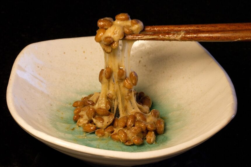
Dietary K1 sufficiency is easy (one salad); dietary K2 is not — menaquinones concentrate in foods most modern diets have marginalised: natto above all, aged cheeses, certain fermented products. This dietary reality, plus the pharmacokinetic case, is why K2 — specifically long-chain MK-7 in its active all-trans form (see the isomer question) — is the rational supplemental form for bone and vascular Gla-protein support, taken with fat like every K vitamin. And one safety note stands above all of it: anyone on warfarin-class anticoagulants must read the interaction rules before any vitamin K product.
The clinical bottom line
K1 and K2 run the same activating chemistry for different clients: K1 funds the liver’s clotting factory; long-chain K2 stays in circulation long enough to activate osteocalcin in bone and MGP in artery walls. Elevated uncarboxylated Gla proteins across populations show the periphery routinely runs under-served — and the Rotterdam data tie higher menaquinone intake specifically to less vascular calcification. Different postcode, different job: that is the K2 story in one line.
Primary sources
- Shearer MJ, Newman P. Metabolism and cell biology of vitamin K. Thromb Haemost. 2008;100(4):530–547. doi:10.1160/TH08-03-0147
- Schurgers LJ, Vermeer C, et al. Vitamin K-containing dietary supplements: comparison of synthetic vitamin K1 and natto-derived menaquinone-7. Thromb Haemost. 2008;100(4):593–603. doi:10.1160/TH08-07-0481
- Geleijnse JM, Vermeer C, Grobbee DE, et al. Dietary intake of menaquinone is associated with a reduced risk of coronary heart disease: the Rotterdam Study. J Nutr. 2004;134(11):3100–3105. doi:10.1093/jn/134.11.3100
- Schurgers LJ, Cranenburg EC, Vermeer C. Matrix Gla-protein: the calcification inhibitor in need of vitamin K. Thromb Haemost. 2008;100(4):593–603 (MGP mechanism sections). doi:10.1160/TH08-07-0481
- Beulens JW, et al. High dietary menaquinone intake is associated with reduced coronary calcification. Atherosclerosis. 2009;203(2):489–493. doi:10.1016/j.atherosclerosis.2008.07.010
Educational information on nutrient function, not medical advice. Patients on anticoagulant therapy must consult their physician before any vitamin K intake change.
Was this article helpful?
Related Coverage


Editorial standards. This article was produced under the GMJ News editorial process, with oversight by the GMJ Editorial Board. Our editorial process. Spotted an error? Contact the editorial team.