By using this site, you agree to the Privacy Policy and Terms of Use.
Accept
GMJ NewsGMJ NewsGMJ News
  • Latest News
    • GMJ Briefs
  • Podcast & Media
    • Podcast Episodes
    • GMJ Audio
    • GMJ Videos
  • Research Digest
    • New Studies
    • Georgian Research
    • Data & Numbers
  • Policy & Systems
    • Health Policy
    • Quality & Safety
    • Migration & Health
    • Global Health
  • Practice
    • Clinical Updates
    • Case Discussions
    • Pharmacy & Prescribing
    • Ingredients A-Z
  • Perspectives
    • Editorial
    • Explainers
    • Voices
    • Letters
  • GMJ Articles
    • Vol. 1 Issue 2 (2026)
    • Vol. 1 Issue 1 (2026)
    • Pre-Launch Articles (2025)
  • Read the Journal →
  • About GMJ News
Notification Show More
Font ResizerAa
GMJ NewsGMJ News
Font ResizerAa
  • Latest News
    • GMJ Briefs
  • Podcast & Media
    • Podcast Episodes
    • GMJ Audio
    • GMJ Videos
  • Research Digest
    • New Studies
    • Georgian Research
    • Data & Numbers
  • Policy & Systems
    • Health Policy
    • Quality & Safety
    • Migration & Health
    • Global Health
  • Practice
    • Clinical Updates
    • Case Discussions
    • Pharmacy & Prescribing
    • Ingredients A-Z
  • Perspectives
    • Editorial
    • Explainers
    • Voices
    • Letters
  • GMJ Articles
    • Vol. 1 Issue 2 (2026)
    • Vol. 1 Issue 1 (2026)
    • Pre-Launch Articles (2025)
  • Read the Journal →
  • About GMJ News
Follow US
GMJ News > Practice > Clinical Updates > New PET Imaging Platform Transforms Real-Time Surgical Margin Assessment in Osteosarcoma
Clinical UpdatesNew StudiesPracticeResearch Digest

New PET Imaging Platform Transforms Real-Time Surgical Margin Assessment in Osteosarcoma

GMJ
Last updated: 07/06/2026 20:42
By
GMJ Practice Desk
Share
6 Min Read
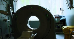
Advanced PET imaging system providing real-time surgical guidance for osteosarcoma tumor resection
Revolutionary PET imaging platform enables real-time tumor margin assessment during osteosarcoma surgery, potentially reducing recurrence risk while preserving limb function. Technology presented at SNMMI 2026 could transform surgical precision in bone cancer treatment. — Photo: Accuray / Pexels
SHARE
🎧 Listen to this article5:03 min · 720 words · GMJ Audio

Updated 07/06/2026

Contents
    • Key takeaways
      • Study at a Glance
  • Revolutionary Real-Time Tumor Detection
  • Precision Surgery with Enhanced Outcomes
  • Implications for Limb-Sparing Surgery
    • What this means
  • Frequently asked questions
    • How does this PET imaging platform differ from current surgical guidance methods?
    • What are the potential benefits for osteosarcoma patients?
    • When might this technology become available in clinical practice?
4 min read|720 words
✓ Medically reviewed by Prof. Giorgi Pkhakadze, MD, MPH, PhD · ORCID 0000-0001-7609-4515

🟡 Preliminary Evidence

A breakthrough end-to-end PET imaging approach for osteosarcoma can rapidly distinguish tumor tissue from normal tissue and accurately assess surgical margins in real time, according to research presented at the Society of Nuclear Medicine and Molecular Imaging 2026 Annual Meeting. The novel platform has the potential to fundamentally reshape surgical practice for osteosarcoma by enabling precise tumor resection and significantly reducing the risk of local recurrence.

Key takeaways

  • New PET imaging platform can rapidly distinguish tumor tissue from normal tissue in real time during surgery
  • Technology accurately assesses surgical margins, potentially reducing local recurrence risk
  • Platform preserves maximum limb function by enabling more precise tumor resection

Study at a Glance

Source SNMMI 2026 Annual Meeting
Study type Platform development
Target condition Osteosarcoma
Technology Integrated PET imaging
Application Real-time surgical guidance
Real-time
surgical margin assessment capability of new PET imaging platform

Revolutionary Real-Time Tumor Detection

The integrated PET imaging platform represents a significant advancement in surgical oncology, offering surgeons real-time visualization of tumor boundaries during osteosarcoma resection. The platform provides immediate molecular-level distinction between malignant and healthy tissue, according to research presented at the SNMMI meeting.

Submit Your Paper
GMJ_Submit_Banner

The technology demonstrates the platform’s ability to rapidly process imaging data and provide clear guidance to surgical teams. This capability addresses a critical challenge in osteosarcoma surgery, where achieving complete tumor removal while preserving maximum limb function remains a delicate balance. For related advances in surgical imaging, see our clinical updates section.

🎙️ Related Podcast Episodes
🎧 #35 | GMJ Podcast | Lung Adenocarcinoma and Tuberculous Lymphadenitis — Diagnostic Challenges in a Rare Case · 19m
🎧 #24 | WHO Releases Updated Health Inequality Data Repository and Equity Toolkit · 21m

Precision Surgery with Enhanced Outcomes

The new platform’s ability to accurately assess surgical margins in real time could significantly reduce the risk of local recurrence, according to the Society of Nuclear Medicine and Molecular Imaging 2026 Annual Meeting presentation. The PET imaging system enables surgeons to make informed decisions about the extent of resection needed while the patient remains on the operating table.

This real-time capability represents a potential improvement over conventional histopathological assessment methods, which may not provide complete margin evaluation until after surgery is completed.

Implications for Limb-Sparing Surgery

The technology’s precision in distinguishing tumor from normal tissue has particular significance for limb-sparing procedures, where preserving functional anatomy while achieving oncological clearance is important. The National Cancer Institute has emphasized the importance of achieving negative surgical margins in osteosarcoma treatment while maintaining quality of life through limb preservation.

This new imaging platform could potentially advance both objectives simultaneously by providing surgeons with molecular-level precision in defining resection boundaries. Our latest research continues to track developments in precision surgical techniques.

The integrated PET imaging platform can rapidly and reliably distinguish tumor tissue from normal tissue and accurately assess surgical margins in real time during osteosarcoma surgery.

— Research findings, Society of Nuclear Medicine and Molecular Imaging (2026 Annual Meeting)

What this means

For patients: Potentially improved surgical outcomes with better margin clearance and enhanced limb preservation, leading to reduced recurrence risk and better functional outcomes
For clinicians: Real-time molecular imaging guidance during surgery enables more precise tumor resection decisions and immediate margin assessment without waiting for histopathological results
For policymakers: Investment in advanced surgical imaging technologies could improve osteosarcoma treatment outcomes

Frequently asked questions

How does this PET imaging platform differ from current surgical guidance methods?

According to the SNMMI 2026 presentation, unlike traditional frozen section analysis that provides histopathological assessment post-resection, this integrated platform offers real-time molecular imaging that distinguishes tumor from normal tissue during active surgery. This enables immediate surgical decision-making rather than retrospective margin evaluation.

What are the potential benefits for osteosarcoma patients?

Based on the research presented, the platform could potentially reduce local recurrence risk by ensuring complete tumor removal while preserving maximum limb function through precise margin definition. This dual benefit addresses two primary concerns in osteosarcoma treatment: oncological clearance and functional preservation.

When might this technology become available in clinical practice?

While presented at the 2026 SNMMI meeting, the timeline for clinical implementation will depend on further validation studies, regulatory approval processes, and institutional adoption. The preliminary evidence suggests promising potential for transforming surgical practice.

The development of this integrated PET imaging platform represents a significant step forward in precision surgical oncology, particularly for complex cases like osteosarcoma where achieving optimal outcomes requires balancing complete tumor removal with functional preservation. As this technology undergoes further validation and potential regulatory review, it could establish new standards for real-time surgical guidance in musculoskeletal oncology.

Source: Integrated PET imaging platform rapidly defines clear margins to guide surgical resection in osteosarcoma

Was this article helpful?

Disclaimer. This article is health journalism intended for general information and education. It is not medical advice and is not a substitute for professional diagnosis or treatment. Always consult a qualified healthcare provider about your individual circumstances. Full disclaimer →

Related Coverage
How Creatine Supplements Reshape Your Body's Methylation CycleJul 21, 2026
Why vitamin D prescriptions favour weekly boluses over daily doses — and what the evidence saysJul 21, 2026
Mitochondria as metabolic decision-makers: how cellular power plants regulate inflammation, aging, and diseaseJul 21, 2026
Stress impairs memory and attention by up to 20%, study finds—but botanical interventions show promiseJul 21, 2026
Related reference
  • Osteosarcoma · Condition
PG
Written by
Prof. Giorgi Pkhakadze, MD, MPH, PhD
Editor-in-Chief, GMJ News
Full profile →  ·  ORCID 0000-0001-7609-4515
Medical disclaimer. This article is health journalism intended for general information. It is not medical advice and is not a substitute for consultation with a qualified healthcare professional. Always seek your physician's advice regarding any medical condition.
Medically reviewed by Prof. Giorgi Pkhakadze, MD, MPH, PhD. Spotted an error? Contact the editorial team.

Get the GMJ News digest
Evidence-based health journalism in your inbox. No spam; unsubscribe anytime.
TAGGED:osteosarcomaPET imagingprecision surgeryreal-time imagingsurgical margins
Share This Article
Facebook LinkedIn Bluesky Copy Link Print
GMJ
ByGMJ Practice Desk
Follow:
GMJ Practice Desk is part of GMJ News, the newsroom of the Georgian Medical Journal (gmj.ge), published by the Public Health Institute of Georgia. Every article is editorially reviewed before publication.
Leave a Comment Leave a Comment

Leave a Reply Cancel reply

Your email address will not be published. Required fields are marked *

Submit Your Paper →

Georgia's peer-reviewed open-access medical journal. No APC until January 2027.
Submit Manuscript →
How Creatine Supplements Reshape Your Body’s Methylation Cycle

Your body produces 2 grams of creatine daily, consuming 40% of your…

Why vitamin D prescriptions favour weekly boluses over daily doses — and what the evidence says

Vitamin D prescriptions typically specify 50,000 IU weekly, a pattern driven by…

Mitochondria as metabolic decision-makers: how cellular power plants regulate inflammation, aging, and disease

Mitochondria function as metabolic control centers that regulate inflammation, aging, and disease…

Submit Your Paper to GMJ

No APC until January 2027.
Submit Manuscript →

You Might Also Like

Medical illustration showing myelin sheath damage from vitamin B12 deficiency
New Studies

B12 Deficiency Damages Nerves Before Blood Tests Show Abnormalities, Studies Find

By
GMJ Research Desk
21/05/2026
Microscopic image of Echinococcus multilocularis tapeworm parasiteIllustrative image · Photo by fr0ggy5 on Unsplash (Unsplash License)
Clinical UpdatesGlobal HealthNew StudiesPolicy & SystemsPracticeResearch Digest

Fatal Tapeworm Disease Spreads to Pacific Northwest Wild Animals for First Time

By
GMJ Practice Desk
07/07/2026
Comparison chart showing treatment response of bradykinin vs histamine-mediated angioedema to standard medicationsIllustrative image · More than makeup- Surgeon Cell Soldier turns moulage art into operational readiness (9245265).jpg by U.S. Army photo by Spc. Micheala Maldonado / Public domain via Wikimedia Commons (Public domain)
Pharmacy & PrescribingPolicy & SystemsPracticeQuality & Safety

ACE Inhibitors and Angioedema: Why Distinguishing Drug Types Matters for Treatment

By
GMJ Practice Desk
10/07/2026
Diagram showing inflammatory reflex pathway from brain through vagus nerve to spleen and immune response
New StudiesResearch Digest

Brain-Immune Connection: How the Inflammatory Reflex Controls Disease

By
GMJ Research Desk
22/05/2026
Facebook Twitter Youtube Instagram
Company
  • Privacy Policy
  • Contact US
  • GMJ Journal
  • Submit Manuscript
  • Editorial Team
  • Register at GMJ
  • Terms of Use

Subscribe to GMJ News — Click here

Join Community
© 2026 Georgian Medical Journal (GMJ). Published by the Public Health Institute of Georgia (PHIG). All rights reserved.
Welcome Back!

Sign in to your account

Username or Email Address
Password

Lost your password?

Not a member? Sign Up