Updated 13/09/2026
🟠 Moderate Evidence
The human brain is far more than a collection of neurons firing in isolation. According to research published in Science Translational Medicine (DOI: 10.1126/scitranslmed.adi7828), the organ’s health and function depend critically on dynamic intercellular communication between multiple cell types—astrocytes, microglia, oligodendrocytes, and neurons—whose molecular conversations determine whether the brain environment becomes protective or inflammatory. These cellular interactions fundamentally shape memory formation, mood regulation, cognitive function, and long-term neural resilience.
Key takeaways
- Astrocytes function as a central regulatory hub, deciding whether the brain environment becomes supportive or pro-inflammatory through signaling molecules
- Microglia can shift between protective and damaging states depending on signals they receive, releasing either anti-inflammatory or pro-inflammatory factors
- Neurons are vulnerable to inflammatory cascade effects including oxidative stress, impaired glutamate handling, and reduced metabolic support
- Oligodendrocytes respond to environmental cues, with inflammatory signals suppressing myelin formation and regenerative signals promoting neuronal stability
The Brain’s Cellular Conversation Network
How four major brain cell types communicate to create protective or inflammatory environments

Adapted from Science Translational Medicine (DOI: 10.1126/scitranslmed.adi7828) | Georgian Medical Journal News
Astrocytes: The Brain’s Central Regulatory Hub
Astrocytes occupy a uniquely powerful position in brain physiology. According to the Science Translational Medicine research, when astrocytes sense danger signals—such as those accompanying stress, illness, or injury—they activate nuclear factor-kappa B (NF-κB), a key transcription factor that orchestrates inflammatory gene expression. This activation triggers the release of pro-inflammatory molecules that reshape the entire brain microenvironment.
Conversely, when conditions are safe and the brain senses security signals, astrocytes shift their behavior dramatically. They release growth factors and neurotrophic molecules that encourage neuron survival and maturation of oligodendrocyte precursor cells. This capacity to toggle between protective and inflammatory states makes astrocytes the brain’s central switchboard, determining whether the neural tissue receives support or faces inflammatory assault. Understanding astrocyte behaviour is foundational to grasping how mood, memory, and resilience are neurobiologically constructed.
Astrocytes decide whether the brain environment becomes supportive or inflammatory. When they sense danger signals, they activate NF-κB and release pro-inflammatory molecules. When conditions are safe, they release factors that help neurons grow and support new oligodendrocyte maturation.
— Research published in Science Translational Medicine, 2024
Microglia: Immune Sentinels With Dual Capability
Microglia are the brain’s resident immune cells, constantly surveilling the neural microenvironment. According to the published research, microglia possess a remarkable capacity for functional plasticity. They can shift between a pro-inflammatory state—in which they release interleukin-1 beta (IL-1β), granulocyte-macrophage colony-stimulating factor (GM-CSF), and other pro-inflammatory cytokines—and a neuroprotective state characterized by the release of anti-inflammatory and growth-supporting molecules.
The critical determinant of this switch is the signals microglia receive from astrocytes and neurons. This represents a crucial insight into brain resilience: microglial activation is not inherently pathogenic. Rather, microglia respond contextually to their chemical environment. In the presence of sustained pro-inflammatory astrocyte signaling, microglia become primed toward a damaging phenotype. However, when astrocytes and neurons communicate safety and regenerative signals, microglia shift to a protective, supportive role. This dynamic reveals why chronic stress and persistent inflammatory signals can lock microglia into a harmful state, while recovery and healing can reprogram their function.
The Neuronal Toll: Vulnerability in the Inflammatory Cascade
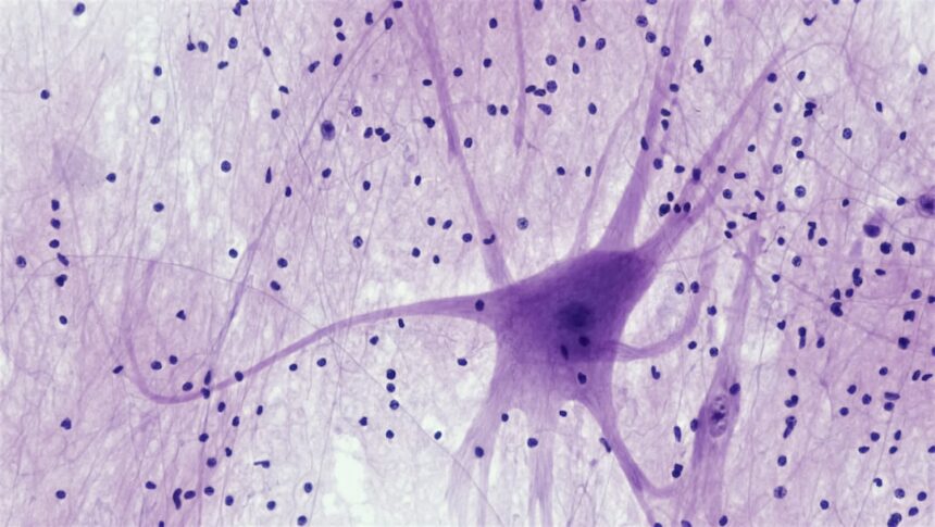
Neurons sit at the centre of this cellular dialogue, yet they are among the most vulnerable participants. When astrocytes and microglia shift toward pro-inflammatory signaling, neurons face a converging assault of metabolic and oxidative insults. According to the research literature, inflammatory conditions impair neurons through multiple mechanisms: elevated oxidative stress damages cell membranes and mitochondria; excessive nitric oxide production creates nitrosative stress; and withdrawal of metabolic support—particularly glucose and lactate provided by astrocytes—forces neurons into bioenergetic crisis.
Perhaps most insidiously, inflammation impairs the neuronal machinery that handles glutamate, the brain’s primary excitatory neurotransmitter. This breakdown in glutamate homeostasis triggers excitotoxicity—a cascade in which excessive glutamate overstimulates neuronal receptors, causing calcium influx, mitochondrial dysfunction, and ultimately cell death. This explains why sustained brain inflammation is linked to cognitive decline, memory impairment, and mood disturbance. The neuron’s fate is not determined solely by its own genes, but by the molecular environment created by its neighbours. See our clinical updates section for more on neuroinflammation and clinical outcomes.
Oligodendrocytes and Myelin: Regeneration Depends on Environmental Cues
Oligodendrocytes are the brain’s myelin-producing cells, responsible for insulating axons and enabling rapid neuronal communication. Oligodendrocyte precursor cells (OPCs) continuously mature into mature oligodendrocytes throughout life, supporting ongoing myelin repair and neural plasticity. However, this regenerative capacity is exquisitely sensitive to environmental signals. According to the published data, pro-inflammatory signals from activated astrocytes and microglia directly suppress OPC maturation and myelin synthesis. Conversely, when astrocytes release regenerative signaling molecules—such as growth factors—OPCs differentiate more readily, myelin formation accelerates, and neuronal stability improves.
This reveals a fundamental principle of brain health: resilience is not a fixed property but an outcome of cellular coordination. Chronic inflammation suppresses the brain’s own repair machinery, locking it into a state of declining myelin and neuronal support. Conversely, recovery from stress or illness reactivates this regenerative network. Understanding that myelin regeneration responds to environmental signals offers a conceptual foundation for why interventions targeting inflammation—whether through lifestyle, pharmacotherapy, or psychosocial approaches—can restore cognitive and emotional health. Visit our new studies section for emerging research on neuroprotection.
What this means
Frequently asked questions
What is the difference between neurons and glia?
Neurons transmit electrical and chemical signals that encode information and drive behaviour. Glia—including astrocytes, microglia, and oligodendrocytes—provide structural support, regulate the neurochemical environment, clear cellular debris, and produce myelin. While neurons are often portrayed as the brain’s “thinking” cells, glial cells actively regulate neuronal function and determine whether the brain environment supports or undermines cognitive and emotional health.
Can chronic inflammation in the brain be reversed?
Yes, emerging evidence suggests that neuroimmune dysfunction can be partially or substantially reversed. Recovery depends on reducing the signals that drive astrocyte and microglial activation—through stress reduction, management of systemic inflammation, improved sleep, metabolic health, and in some cases, targeted anti-inflammatory interventions. The brain possesses intrinsic repair capacity, but this capacity is suppressed when pro-inflammatory signaling persists.
How do lifestyle factors like sleep and exercise influence these brain cell conversations?
Sleep and physical activity directly modulate astrocyte and microglial signaling. During sleep, astrocytes orchestrate the glymphatic system, which clears metabolic waste and supports synaptic plasticity. Exercise shifts microglia toward neuroprotective phenotypes and enhances astrocyte-mediated metabolic support for neurons. Chronic sleep deprivation and sedentary behaviour, conversely, promote pro-inflammatory microglial states and impair astrocyte function, accelerating cognitive decline.
The emerging field of neuroimmunology has fundamentally reshaped our understanding of brain health. Rather than viewing the brain as a collection of independent neurons, we now recognize it as a sophisticated cellular ecosystem in which constant communication between multiple cell types determines whether the organ becomes resilient or vulnerable. As research continues to map these interactions and identify interventions that restore healthy glial signaling, the potential for new preventive and therapeutic approaches to neuropsychiatric and neurodegenerative disease expands significantly.
Source: Research on glial-neuronal communication in brain stress and inflammation, Science Translational Medicine, 2024
🗺️ Find certified Georgian healthcare providers: sheniekimi.ge — Georgia Health Map · 998 clinics · 6,600+ doctors · 2,296 pharmacies
Was this article helpful?
Disclaimer. This article is health journalism intended for general information and education. It is not medical advice and is not a substitute for professional diagnosis or treatment. Always consult a qualified healthcare provider about your individual circumstances. Full disclaimer →
Related Coverage




Editorial standards. This article was produced under the GMJ News editorial process, with oversight by the GMJ Editorial Board. Our editorial process. Spotted an error? Contact the editorial team.