
Updated 13/09/2026
🟠 Moderate Evidence
Docosahexaenoic acid (DHA), a 22-carbon omega-3 fatty acid, is not a nutrient that merely “supports” brain function from outside the organ. Rather, DHA is a structural component of the brain itself—constituting approximately 40% of all polyunsaturated fatty acids in the brain and over 90% of the brain’s omega-3 content, according to biochemical analysis cited in the neuroscience literature. This distinction fundamentally reframes how scientists and clinicians should understand omega-3 fatty acids and brain health.
Key takeaways
- DHA comprises 40% of brain polyunsaturated fatty acids and over 90% of brain omega-3 content—it is structural, not supplementary
- DHA’s six double bonds create membrane fluidity that determines how quickly neuronal signaling proteins respond to signals
- Patients with major depressive disorder showed 22% lower DHA in the orbitofrontal cortex, with deficits reaching 32% in women versus 16% in men
- DHA deficiency appears region-specific in the brain, suggesting targeted rather than systemic effects
DHA as a Percentage of Brain Lipid Composition
DHA dominates omega-3 fatty acid pools in neural tissue, 2007 neurochemical analysis

Source: Neurochemical literature analysis | Georgian Medical Journal News
DHA as Molecular Architecture, Not Medication
DHA sits primarily at the sn-2 position of phosphatidylethanolamine and phosphatidylserine, two of the dominant phospholipids in neuronal membranes, according to structural lipid biology. Each of its six double bonds introduces a bend in the carbon chain. Those accumulated kinks prevent the tails from packing tightly, which increases membrane fluidity—a physical property that is not abstract or theoretical.
That membrane fluidity directly determines how quickly every receptor, ion channel, and signaling protein embedded in the membrane can change conformation in response to a neural signal. This is the mechanism by which DHA affects brain function: not through a circulating “support” mechanism, but through its role as a foundational structural element of the very cells performing cognition and emotion regulation.
Evidence of Regional DHA Deficiency in Depression
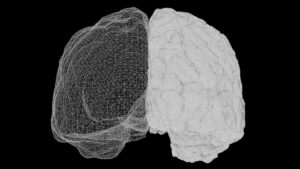
McNamara and colleagues, writing in Biological Psychiatry in 2007, measured the total fatty acid composition of postmortem orbitofrontal cortex tissue (Brodmann Area 10) in 15 patients with Depressive Disorder">major depressive disorder and 27 age-matched controls. After statistical correction for multiple comparisons, DHA was the only fatty acid that differed significantly between groups. Patients with major depressive disorder had 22% less DHA in the orbitofrontal cortex than controls.
The deficit was not uniform across sexes. Women with major depressive disorder showed a 32% reduction in DHA, while men showed a 16% reduction. Importantly, the researchers documented that this deficit could not be wholly attributed to lifestyle factors or postmortem tissue handling variables—suggesting a genuine biological difference in brain DHA content associated with the disorder.
DHA was the only fatty acid that was significantly different between major depressive disorder patients and controls in the orbitofrontal cortex, with patients showing 22% lower levels overall (32% in women, 16% in men).
— McNamara et al., Biological Psychiatry (2007)
Region-Specific Effects and Future Research Directions
An important nuance constrains broad interpretation: the DHA deficit appears to be region-specific. Later studies from the same research group found no significant DHA differences in other brain regions including the amygdala and hippocampus. This regional specificity raises a critical question: is DHA deficiency a marker of regional neuronal dysfunction, or does regional DHA depletion drive regional pathology?
This distinction matters for future research. If DHA deficiency is a primary cause of orbitofrontal cortex dysfunction in depression, DHA supplementation might be therapeutically targeted. If regional DHA depletion is a consequence of underlying neuroinflammation or metabolic dysfunction, supplementation alone may not be sufficient. Ongoing neuroscience research continues to clarify these mechanistic pathways.
What this means
Frequently asked questions
Is DHA supplementation proven to treat depression?
No. While evidence from Biological Psychiatry shows that patients with depression have lower DHA in specific brain regions, this is an association, not proof of causation. Clinical trials of DHA supplementation for depression have shown mixed results and do not yet support DHA as a first-line treatment. The orbitofrontal cortex finding is important for understanding disease biology, but does not automatically translate to therapeutic benefit.
Does the brain’s high DHA content mean I need to supplement?
Not necessarily. The brain’s structural dependence on DHA means that adequate dietary DHA is essential—but for most people consuming fish, seafood, or plant-based omega-3 sources, dietary intake is sufficient. Supplementation may be warranted in vegans, people unable to consume fish, or those with documented absorption disorders. Individual assessment by a clinician is preferable to universal supplementation.
Why is the DHA deficit in women with depression larger than in men?
The reason for sex-specific differences (32% in women versus 16% in men) remains unclear. Possibilities include differences in DHA absorption, metabolism, or utilization between sexes; differences in how depression manifests neurobiologically; or differences in hormonal factors affecting lipid metabolism. This observation warrants further investigation but does not yet support sex-specific treatment recommendations.
Understanding DHA as structural rather than supplementary invites a more rigorous conversation about brain health. Rather than asking whether omega-3 supplements can fix cognitive or emotional problems, researchers now ask: what are the consequences of suboptimal DHA during critical periods of brain development, and how does DHA insufficiency contribute to specific regional pathology in psychiatric illness? Future clinical research will determine whether correcting DHA deficiency in specific populations produces measurable improvements in symptoms or outcomes.
Source: McNamara et al., Biological Psychiatry (2007)
🗺️ Find certified Georgian healthcare providers: sheniekimi.ge — Georgia Health Map · 998 clinics · 6,600+ doctors · 2,296 pharmacies
Was this article helpful?
Disclaimer. This article is health journalism intended for general information and education. It is not medical advice and is not a substitute for professional diagnosis or treatment. Always consult a qualified healthcare provider about your individual circumstances. Full disclaimer →
Related Coverage


Editorial standards. This article was produced under the GMJ News editorial process, with oversight by the GMJ Editorial Board. Our editorial process. Spotted an error? Contact the editorial team.