By using this site, you agree to the Privacy Policy and Terms of Use.
Accept
GMJ NewsGMJ NewsGMJ News
  • Latest News
    • GMJ Briefs
  • Podcast & Media
    • Podcast Episodes
    • GMJ Audio
    • GMJ Videos
  • Research Digest
    • New Studies
    • Georgian Research
    • Data & Numbers
  • Policy & Systems
    • Health Policy
    • Quality & Safety
    • Migration & Health
    • Global Health
  • Practice
    • Clinical Updates
    • Case Discussions
    • Pharmacy & Prescribing
    • Ingredients A-Z
  • Perspectives
    • Editorial
    • Explainers
    • Voices
    • Letters
  • GMJ Articles
    • Vol. 1 Issue 2 (2026)
    • Vol. 1 Issue 1 (2026)
    • Pre-Launch Articles (2025)
  • Read the Journal →
  • About GMJ News
Notification Show More
Font ResizerAa
GMJ NewsGMJ News
Font ResizerAa
  • Latest News
    • GMJ Briefs
  • Podcast & Media
    • Podcast Episodes
    • GMJ Audio
    • GMJ Videos
  • Research Digest
    • New Studies
    • Georgian Research
    • Data & Numbers
  • Policy & Systems
    • Health Policy
    • Quality & Safety
    • Migration & Health
    • Global Health
  • Practice
    • Clinical Updates
    • Case Discussions
    • Pharmacy & Prescribing
    • Ingredients A-Z
  • Perspectives
    • Editorial
    • Explainers
    • Voices
    • Letters
  • GMJ Articles
    • Vol. 1 Issue 2 (2026)
    • Vol. 1 Issue 1 (2026)
    • Pre-Launch Articles (2025)
  • Read the Journal →
  • About GMJ News
Follow US
GMJ News > Perspectives > Explainers > How Cells Move Materials: The Microtubule Transport System Explained
ExplainersPerspectives

How Cells Move Materials: The Microtubule Transport System Explained

GMJ
Last updated: 12/07/2026 13:29
By
GMJ Perspectives Desk
Share
7 Min Read
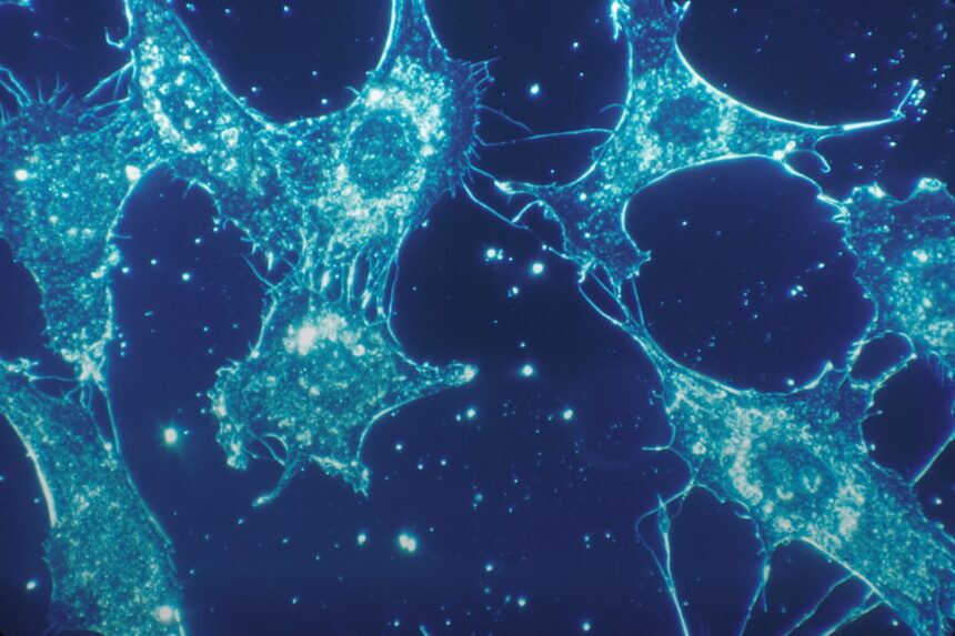
Glowing microtubule filaments showing dynamic assembly and disassembly inside a living cell under fluorescence microscopyIllustrative image · Photo by National Cancer Institute on Unsplash (Unsplash License)
Microtubules—cylindrical protein structures inside cells—continuously rebuild themselves through dynamic instability, enabling cell division, neuronal transport over meter-long distances, and cellular migration. Understanding this system reveals both how cancer drugs work and what goes wrong in neurodegenerative diseases. — Photo by National Cancer Institute on Unsplash (Unsplash License)
SHARE
5 min read|962 words
✓ Reviewed by Prof. Giorgi Pkhakadze, MD, MPH, PhD · ORCID 0000-0001-7609-4515

Inside every living cell, a continuous reorganization of protein structures enables the transport of materials, signal propagation, and cellular structure maintenance. Microtubules—cylindrical protein polymers composed primarily of α- and β-tubulin dimers—are rebuilt constantly through a process called dynamic instability, allowing cells to adapt their internal architecture in real time without forming permanent tracks.

Contents
    • Key takeaways
      • Microtubule Functions in Cellular Processes
  • The Continuous Rebuilding Process Behind Cellular Function
  • Therapeutic Targeting and Disease Mechanisms
  • Clinical Implications Across Medicine and Research
    • What this means
  • Frequently asked questions
    • What is dynamic instability and why does it matter?
    • How do cancer drugs target microtubules?
    • Why does Alzheimer’s disease involve microtubules?

Key takeaways

  • Microtubules continuously assemble and disassemble through dynamic instability, rebuilding rather than remaining static
  • This transport system is essential for accurate cell division, neuronal function over distances exceeding one meter, and cellular migration during development
  • Disruption of microtubule dynamics underlies both therapeutic cancer interventions and pathological processes in neurodegenerative disease
1 metre
Maximum distance some neurons span, all dependent on functional microtubule transport systems for intracellular delivery

Microtubule Functions in Cellular Processes

Key cellular processes dependent on microtubule dynamics and transport

Mitotic spindle formation
Critical
Neuronal transport
Critical
Cell migration
Critical
Organelle positioning
Essential
Ciliary/flagellar motility

Specialized

Source: Cell Biology Literature | Georgian Medical Journal News

The Continuous Rebuilding Process Behind Cellular Function

Microtubule dynamic instability was first characterized as a stochastic process in which individual microtubules undergo phases of growth and shrinkage rather than stable polymerization. This fundamental property, observed through live-cell video microscopy as glowing streaks marking the growing ends of microtubules, represents a departure from earlier static models of cellular architecture.

Submit Your Paper
GMJ_Submit_Banner

The continuous assembly and disassembly cycle occurs through the sequential binding and hydrolysis of guanosine triphosphate (GTP) at the microtubule plus end. This allows cells to reorganize their internal cytoskeletal layout in response to developmental cues, external signals, and functional demands—a flexibility that would be impossible if microtubules formed permanent tracks.

🎙️ Related Podcast Episodes
🎧 #39 | GMJ Podcast | Acne and Metabolic Dysfunction — Insulin Resistance, IGF-1, and Clinical Implications · 15m
🎧 #38 | GMJ Podcast | Acne and Metabolic Dysfunction — Insulin Resistance, IGF-1, and Clinical Implications · 21m
🎧 #27 | WHO Calls for Environmentally Friendly and Less Invasive Oral Health Care · 21m

Therapeutic Targeting and Disease Mechanisms

Cancer chemotherapy agents such as taxanes (docetaxel, paclitaxel) and vinca alkaloids work by either stabilizing microtubules in a polymerized state or inducing complete depolymerization, thereby preventing the mitotic spindle formation required for cell division. This mechanism exploits the fact that rapidly dividing cancer cells are acutely dependent on functional microtubule dynamics.

Conversely, failures in microtubule-associated protein function—particularly tau hyperphosphorylation and aggregation—disrupt the stability and transport capacity of microtubules in neurons, leading to impaired axonal transport and intracellular accumulation of toxic proteins characteristic of Alzheimer’s disease and other tauopathies. These pathological processes illustrate how precise microtubule regulation is essential for neuronal survival over the lifespan.

Clinical Implications Across Medicine and Research

The understanding of microtubule biology has expanded beyond cancer therapeutics. Structural studies using cryo-electron microscopy have revealed the atomic-level architecture of the tubulin dimer and microtubule lattice, enabling rational drug design targeting specific aspects of microtubule dynamics. Current research is exploring selective modulators of microtubule stability that may offer therapeutic benefit in neurodegenerative diseases while minimizing toxicity to non-dividing cells.

In developmental biology and tissue repair, controlled modulation of microtubule dynamics drives cell migration and establishment of cellular polarity—critical processes in embryonic development, wound healing, and immune cell function. Dysregulation of these processes is implicated in birth defects, impaired wound healing, and metastatic cancer progression.

Microtubule dynamic instability—the continuous cycling between growth and shrinkage—is not a cellular inefficiency but rather a fundamental design principle that enables rapid cellular adaptation, accurate division, and long-distance transport in specialized cell types such as neurons.

— Cell Biology Research Community, Multiple Institutions (Nature Reviews Molecular Cell Biology and related literature)

What this means

For patients: Understanding that microtubule dysfunction underlies both cancer and neurodegeneration offers hope for improved therapies. Patients receiving taxane-based chemotherapy are experiencing drugs that exploit this cellular system; those with Alzheimer’s disease may benefit from emerging therapeutic approaches that restore microtubule stability and axonal transport.
For clinicians: Microtubule-targeting drugs remain cornerstone cancer therapies, but emerging evidence suggests that selective stabilization of microtubules may benefit neurodegenerative disease patients—requiring careful patient selection and monitoring for off-target effects on rapidly dividing cells.
For policymakers: Investment in basic cell biology research continues to yield clinically transformative insights. Funding for structural biology, drug discovery targeting microtubule regulators, and biomarker development for patient stratification can accelerate translation of these discoveries into improved therapeutics.

Frequently asked questions

What is dynamic instability and why does it matter?

Dynamic instability is the inherent property of microtubules to randomly switch between phases of polymerization (growth) and depolymerization (shrinkage). Rather than being a flaw, this feature allows cells to rapidly reorganize their internal structure without constructing permanent scaffolding. This adaptability is essential for cell division, migration, and neuronal function, making it one of the most fundamental mechanisms in cell biology.

How do cancer drugs target microtubules?

Cancer chemotherapy agents such as paclitaxel (Taxol) and docetaxel stabilize microtubules by preventing their disassembly, thereby blocking the formation of the mitotic spindle required for cell division. Other agents like vincristine induce microtubule collapse. These drugs exploit the fact that cancer cells divide rapidly and are thus more sensitive to disruption of microtubule dynamics than most normal tissues, though toxicity to dividing cells in bone marrow and the gastrointestinal tract remains a significant clinical challenge.

Why does Alzheimer’s disease involve microtubules?

In Alzheimer’s disease, the protein tau—which normally stabilizes microtubules and is essential for axonal transport—becomes abnormally phosphorylated and forms aggregates. This causes microtubules to become unstable and non-functional, impairing the transport of nutrients and signaling molecules along neuronal axons that can span over a meter in length. The resulting breakdown of intracellular transport leads to neuronal death and cognitive decline.

Research into microtubule biology continues to advance rapidly, with new imaging techniques revealing real-time dynamics at unprecedented resolution and with novel drug candidates designed to selectively modulate microtubule stability for therapeutic benefit. Understanding this fundamental cellular system remains central to developing treatments for cancer, neurodegenerative disease, and developmental disorders. For more information on cellular biology and disease mechanisms, explore our latest research updates and in-depth explainers on cell biology.

Source: Cell Biology Visualization and Microtubule Dynamics | Video Credit: Andy Moore

Was this article helpful?

Disclaimer. This article is health journalism intended for general information and education. It is not medical advice and is not a substitute for professional diagnosis or treatment. Always consult a qualified healthcare provider about your individual circumstances. Full disclaimer →

Related Coverage

Beyond Minimum Requirements: New Evidence Suggests Higher Protein Intake Supports Healthy AgingAug 5, 2026
Five Foods Cardiologists Recommend for Men's Heart Health: Evidence from Clinical PracticeAug 5, 2026
Understanding UV Radiation: What the Science Says About Sun ProtectionAug 4, 2026
Why Nutrients Work Better Together: How Coordinated Inputs Reshape Brain SynapsesAug 4, 2026
PG
Written by
Prof. Giorgi Pkhakadze, MD, MPH, PhD
Editor-in-Chief, GMJ News
Full profile →  ·  ORCID 0000-0001-7609-4515
Medical disclaimer. This article is health journalism intended for general information. It is not medical advice and is not a substitute for consultation with a qualified healthcare professional. Always seek your physician's advice regarding any medical condition.
Medically reviewed by Prof. Giorgi Pkhakadze, MD, MPH, PhD. Spotted an error? Contact the editorial team.
Get the GMJ News digest
Evidence-based health journalism in your inbox. No spam; unsubscribe anytime.
TAGGED:cancer therapycell-biologycell-dynamicsmicrotubulesneurodegenerative disease
Share This Article
Facebook LinkedIn Bluesky Copy Link Print
GMJ
ByGMJ Perspectives Desk
Follow:
GMJ Perspectives Desk is part of GMJ News, the newsroom of the Georgian Medical Journal (gmj.ge), published by the Public Health Institute of Georgia. Every article is editorially reviewed before publication.
Leave a Comment Leave a Comment

Leave a Reply Cancel reply

Your email address will not be published. Required fields are marked *

Submit Your Paper →

Georgia's peer-reviewed open-access medical journal. No APC until January 2027.
Submit Manuscript →
Waist Circumference Outperforms BMI in Predicting Disability Risk, Whitehall II Cohort Study Shows

A new analysis of the Whitehall II cohort study shows that waist…

FDA-Cleared AI Tool EchoNext Enables ECG-Based Heart Disease Screening

Pathway Labs' EchoNext, an FDA-cleared artificial intelligence tool, enables detection of structural…

Beyond Minimum Requirements: New Evidence Suggests Higher Protein Intake Supports Healthy Aging

A new review of aging and nutrition research suggests that protein intake…

Submit Your Paper to GMJ

No APC until January 2027.
Submit Manuscript →

You Might Also Like

Infographic showing mineral roles in brain function: iodine for thyroid, iron for dopamine synthesis, lithium for neuroplasticity, zinc for synaptic signalingIllustrative image · Photo by Shawn Day on Unsplash (Unsplash License)
Clinical UpdatesExplainersNew StudiesPerspectivesPracticeResearch Digest

How Minerals Shape Brain Function: Evidence from Neuroscience on Micronutrient Deficiency and Cognition

By
GMJ Practice Desk
01/08/2026
Diagram of innate and adaptive immune responses showing macrophages, antibodies, T cells, and immunological memoryIllustrative image · Photo by Vanessa Ray on Pexels (Pexels License)
ExplainersPerspectives

How the immune system learns and remembers: innate defenses meet adaptive immunity

By
GMJ Perspectives Desk
28/07/2026
Diagram showing autonomic nervous system pathways: sympathetic fight-flight versus parasympathetic rest-digest across heart, lungs, digestive system, and pupilsIllustrative image · Photo by Bhautik Patel on Unsplash (Unsplash License)
Clinical UpdatesExplainersPerspectivesPractice

How Your Body Switches Between Two Operating Systems: The Autonomic Nervous System Explained

By
GMJ Practice Desk
26/07/2026
Illustration of creatine's role in mitochondrial ATP production and brain energy in womenIllustrative image · Photo by Andrea Piacquadio on Pexels (Pexels License)
Clinical UpdatesExplainersPerspectivesPractice

Creatine for Women: Beyond Muscle—Evidence for Brain, Energy, and Long-Term Health

By
GMJ Practice Desk
02/08/2026
Facebook Twitter Youtube Instagram
Company
  • Privacy Policy
  • Contact US
  • GMJ Journal
  • Submit Manuscript
  • Editorial Team
  • Register at GMJ
  • Terms of Use

Subscribe to GMJ News — Click here

Join Community
© 2026 Georgian Medical Journal (GMJ). Published by the Public Health Institute of Georgia (PHIG). All rights reserved.
Welcome Back!

Sign in to your account

Username or Email Address
Password

Lost your password?

Not a member? Sign Up