



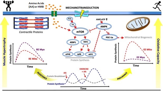
Your cells continuously manufacture new mitochondria in response to increased energy demand, a process called mitochondrial biogenesis that underpins metabolic adaptation to exercise, fasting, and thermal stress. This cellular mechanism involves a coordinated chain of signalling events centred on the master regulator protein PGC-1α (peroxisome proliferator-activated receptor gamma coactivator 1-alpha), which orchestrates both nuclear gene expression and mitochondrial DNA replication.
- The energy sensor cascade: AMPK and SIRT1 trigger mitochondrial growth
- PGC-1α: the master switch coordinating nuclear and mitochondrial programs
- TFAM and mtDNA replication: completing the mitochondrial factory
- Physiological outcomes: enhanced metabolic capacity and cellular resilience
- Frequently asked questions
Key takeaways
- Mitochondrial biogenesis is triggered by energy stress signals including exercise, fasting, cold exposure, and caloric restriction
- The protein PGC-1α acts as the central “master switch”, activated by energy sensors AMPK and SIRT1 when cellular ATP levels decline
- New mitochondria require coordinated synthesis of nuclear-encoded proteins and mitochondrial DNA replication via the transcription factor TFAM
- Increased mitochondrial mass improves cellular energy production capacity and metabolic resilience
Mitochondrial biogenesis: cellular triggers and regulatory pathway
Key signalling molecules and their activation in response to energy demand
Based on exercise physiology and cellular stress response literature | Georgian Medical Journal News
The energy sensor cascade: AMPK and SIRT1 trigger mitochondrial growth
Mitochondrial biogenesis begins with a cellular energy deficit. When ATP (adenosine triphosphate) levels fall during exercise or fasting, two key energy-sensing proteins activate: AMPK (AMP-activated protein kinase) and SIRT1 (a NAD+-dependent deacetylase). Both are sensitive to shifts in the cellular energy charge—specifically, rising AMP and declining NAD+ concentrations signal the need for increased energy production capacity.

AMPK directly phosphorylates PGC-1α, while SIRT1 deacetylates it; both modifications increase PGC-1α activity. This dual activation ensures that only genuine energy stress—not random metabolic noise—triggers the expensive process of building new mitochondria. The redundancy provides robustness to the system, a principle documented across cellular stress response research.
PGC-1α: the master switch coordinating nuclear and mitochondrial programs
Once activated, PGC-1α acts as a transcriptional coactivator, binding to and enhancing the activity of nuclear transcription factors including NRF1 (nuclear respiratory factor 1) and NRF2. Together, these factors increase expression of dozens of nuclear genes encoding mitochondrial proteins—enzymes of the citric acid cycle, components of the electron transport chain, and proteins required for mtDNA replication and repair.
This represents a critical asymmetry in eukaryotic cell biology: although mitochondria have their own small genome (mtDNA), encoding only 13 protein-coding genes, the vast majority of mitochondrial proteins (~1,000 in mammals) are nuclear-encoded, synthesized in the cytoplasm, and imported across the mitochondrial membranes. PGC-1α orchestrates the production of these nuclear-derived proteins, ensuring that mitochondrial biogenesis is balanced and functional. Research published in Current Opinion in Cell Biology has characterised this coordination in detail.
In clinical practice, PGC-1α activation is the mechanistic target of many exercise and dietary interventions aimed at improving mitochondrial function in metabolic diseases such as type 2 diabetes.
TFAM and mtDNA replication: completing the mitochondrial factory
Within mitochondria, a second transcription factor, TFAM (mitochondrial transcription factor A), takes the lead. TFAM binds to mitochondrial DNA and promotes both transcription of the 13 mtDNA-encoded genes and replication of the mtDNA molecule itself. When TFAM levels increase—a consequence of PGC-1α-driven nuclear gene expression—mitochondrial DNA copy number rises, allowing the cell to manufacture more mitochondrial proteins and expand the mitochondrial compartment.
This process is essential for creating functionally mature new mitochondria. Without adequate mtDNA replication and TFAM-driven mtDNA transcription, newly synthesized mitochondrial proteins cannot be assembled into intact, efficient energy-converting organelles. Studies examining exercise adaptation have shown that mtDNA copy number increases proportionally with mitochondrial mass in human skeletal muscle.
Physiological outcomes: enhanced metabolic capacity and cellular resilience
The end result of this coordinated programme is a substantial increase in mitochondrial number, size, and respiratory capacity. In human skeletal muscle, regular aerobic exercise induces a 50–100% increase in mitochondrial volume density within 4–12 weeks, according to exercise physiology literature. This translates to higher oxidative phosphorylation capacity, improved ATP production efficiency, and greater resilience to metabolic stress.
Enhanced mitochondrial function has broad health implications. In public health contexts, increasing mitochondrial biogenesis through physical activity and intermittent fasting is a non-pharmacological strategy for preventing and managing age-related metabolic decline, insulin resistance, and neurodegeneration. The mechanism also explains why sedentary behaviour accelerates mitochondrial decline and why structured exercise interventions can partially reverse age-related metabolic dysfunction.
Mitochondrial biogenesis is a precisely orchestrated cellular programme activated by energy stress, centred on PGC-1α-mediated coordination between nuclear gene expression and mitochondrial DNA replication, resulting in a 50–100% increase in mitochondrial volume density within weeks of regular exercise training.
— Exercise physiology and cellular stress response research
What this means
Frequently asked questions
How quickly do new mitochondria form after starting exercise?
Molecular signals (AMPK, SIRT1, PGC-1α activation) occur within hours of exercise. Increases in mitochondrial protein synthesis begin within 1–2 days. However, detectable increases in total mitochondrial number and oxidative capacity typically require 3–4 weeks of consistent training, as documented in human training studies.
Does fasting trigger mitochondrial biogenesis as effectively as exercise?
Yes. Both exercise and fasting activate AMPK and SIRT1 through energy deficit mechanisms. However, exercise provides additional stimuli (mechanical stress, reactive oxygen species) that may amplify the response. Research suggests that intermittent fasting and caloric restriction activate PGC-1α and increase mitochondrial content, though the magnitude of adaptation is typically smaller than with regular aerobic training.
Can mitochondrial biogenesis be lost if exercise stops?
Yes. Mitochondrial adaptations are highly dynamic and reversible. Cessation of regular exercise leads to a decline in mitochondrial mass and function within 2–4 weeks, a process called mitochondrial detraining. This underscores the importance of sustained physical activity for maintaining metabolic health throughout life.
The discovery of mitochondrial biogenesis as a regulated, trainable process has fundamentally changed how clinicians and researchers approach metabolic disease prevention and treatment. Rather than viewing mitochondrial function as fixed, we now understand it as a plastic trait, responsive to behavioural and environmental cues. This opens pathways for personalised medicine—tailoring exercise, nutritional, and thermal interventions to optimise each individual’s mitochondrial and metabolic capacity. Future research will likely identify additional molecular switches and develop pharmacological tools to enhance mitochondrial biogenesis in ageing and disease contexts.
Source: Mitochondrial biogenesis made “simple”
Was this article helpful?
Disclaimer. This article is health journalism intended for general information and education. It is not medical advice and is not a substitute for professional diagnosis or treatment. Always consult a qualified healthcare provider about your individual circumstances. Full disclaimer →
Related Coverage


Editorial standards. This article was produced under the GMJ News editorial process, with oversight by the GMJ Editorial Board. Our editorial process. Spotted an error? Contact the editorial team.