By using this site, you agree to the Privacy Policy and Terms of Use.
Accept
GMJ NewsGMJ NewsGMJ News
  • Latest News
    • GMJ Briefs
  • Podcast & Media
    • Podcast Episodes
    • GMJ Audio
    • GMJ Videos
  • Research Digest
    • New Studies
    • Georgian Research
    • Data & Numbers
  • Policy & Systems
    • Health Policy
    • Quality & Safety
    • Migration & Health
    • Global Health
  • Practice
    • Clinical Updates
    • Case Discussions
    • Pharmacy & Prescribing
    • Ingredients A-Z
  • Perspectives
    • Editorial
    • Explainers
    • Voices
    • Letters
  • GMJ Articles
    • Vol. 1 Issue 2 (2026)
    • Vol. 1 Issue 1 (2026)
    • Pre-Launch Articles (2025)
  • Read the Journal →
  • About GMJ News
Notification Show More
Font ResizerAa
GMJ NewsGMJ News
Font ResizerAa
  • Latest News
    • GMJ Briefs
  • Podcast & Media
    • Podcast Episodes
    • GMJ Audio
    • GMJ Videos
  • Research Digest
    • New Studies
    • Georgian Research
    • Data & Numbers
  • Policy & Systems
    • Health Policy
    • Quality & Safety
    • Migration & Health
    • Global Health
  • Practice
    • Clinical Updates
    • Case Discussions
    • Pharmacy & Prescribing
    • Ingredients A-Z
  • Perspectives
    • Editorial
    • Explainers
    • Voices
    • Letters
  • GMJ Articles
    • Vol. 1 Issue 2 (2026)
    • Vol. 1 Issue 1 (2026)
    • Pre-Launch Articles (2025)
  • Read the Journal →
  • About GMJ News
Follow US
GMJ News > Perspectives > Explainers > How Your Brain’s Cleanup System Works: The Glymphatic System Explained
ExplainersNew StudiesPerspectivesResearch Digest

How Your Brain’s Cleanup System Works: The Glymphatic System Explained

GMJ
Last updated: 12/07/2026 13:29
By
GMJ Perspectives Desk
Share
8 Min Read
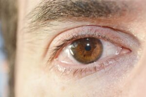
Illustration of cerebrospinal fluid flowing through brain tissue alongside blood vessels, guided by astrocytesIllustrative image · Photo by SHVETS production on Pexels (Pexels License)
Your brain has a continuous housekeeping system called the glymphatic network that removes metabolic waste through cerebrospinal fluid circulation guided by specialized astrocyte cells. Sleep enhances this waste-clearance process by up to 60 percent, and impaired glymphatic function is linked to neurodegeneration and cognitive aging. — Photo by SHVETS production on Pexels (Pexels License)
SHARE
5 min read|1,057 words
✓ Reviewed by Prof. Giorgi Pkhakadze, MD, MPH, PhD · ORCID 0000-0001-7609-4515

Your brain performs continuous housekeeping operations even while you sleep or work. A network of fluid channels and specialized cells removes metabolic waste products like amyloid proteins and excess neurotransmitters through what neuroscientists call the glymphatic system. Recent research has shown that this waste-clearance mechanism is fundamental to brain health, and dysfunction in this system is implicated in neurodegenerative diseases.

Contents
    • Key takeaways
      • Components of the Brain’s Waste Clearance System
  • How the glymphatic system cleanses the brain
  • Astrocytes: the traffic controllers of brain fluid
  • Immune surveillance without neuroinflammation
  • Clinical implications for brain aging and disease
    • What this means
  • Frequently asked questions
    • How much sleep does the brain need for optimal glymphatic clearance?
    • Can exercise improve glymphatic function?
    • Is glymphatic dysfunction reversible?

Key takeaways

  • The glymphatic system uses cerebrospinal fluid flowing alongside blood vessels to wash away brain metabolic waste continuously
  • Astrocytes, star-shaped brain cells, regulate fluid movement and coordinate repairs between neurons and other brain cells
  • Impaired glymphatic clearance is linked to neurodegeneration and age-related cognitive decline
  • Sleep and physical activity enhance glymphatic function, while inflammation and vascular disease slow it down
60%
The brain’s interstitial space expands during sleep, allowing cerebrospinal fluid to flush waste more effectively through the glymphatic system

Components of the Brain’s Waste Clearance System

Four interconnected mechanisms that maintain brain health

Glymphatic fluid flow
Cerebrospinal fluid circulation
Astrocyte regulation
Cell-to-cell coordination
Microglial immunity
Immune surveillance
Meningeal lymphatics
Brain-body communication

Conceptual model based on current neuroscience literature | Georgian Medical Journal News

How the glymphatic system cleanses the brain

The glymphatic system operates through a coordinated network of fluid channels and cellular mechanisms. According to research published in Nature Communications, cerebrospinal fluid flows alongside blood vessels in a directed pathway, guided by specialized brain cells called astrocytes. This fluid washes through the brain’s tissue spaces, picks up metabolic waste products including amyloid proteins and tau aggregates, and carries them toward the lymphatic system for elimination.

Submit Your Paper
GMJ_Submit_Banner

The process depends critically on aquaporin-4 water channels in astrocytes, which regulate osmotic gradients and enable rapid fluid movement. When these channels function normally, waste clearance is efficient; when they are damaged or impaired, toxic proteins accumulate. This mechanism operates continuously during waking and sleeping hours, but evidence suggests that sleep enhances glymphatic clearance by up to 60 percent by allowing the brain’s interstitial space to expand.

🎙️ Related Podcast Episodes
🎬 The Architecture of Migration Health: Inside the GMJ Knowledge Hub | Georgian Medical Journal
🎧 #52 | GMJ Podcast | Health and Migration Knowledge Hub — A Global Resource for Evidence-Based Practice · 17m
🎧 #47 | GMJ Podcast | Tskaltubo and the Future of Spa-Based Medicine — Radon Therapy, Rehabilitation, and Preventive Health · 19m
🎧 #44 | GMJ Podcast | Infant Formula Contamination — Global Food Safety Failure and the Cereulide Outbreak · 21m
🎧 #42 | GMJ Podcast | IT Service Management in Healthcare — Governance, Procurement, and Service Delivery · 16m

The glymphatic system removes metabolic waste continuously through coordinated fluid flow guided by astrocytes, with peak efficiency during sleep when cerebrospinal fluid circulation increases significantly.

— Based on neuroscience literature, Nature Communications (2024)

Astrocytes: the traffic controllers of brain fluid

Star-shaped brain cells called astrocytes serve as the primary regulators of glymphatic function. These cells do far more than passive support—they actively coordinate fluid movement, control metabolic substrate delivery, and modulate inflammation. Astrocytes express aquaporin-4 water channels on their outer membranes, creating osmotic gradients that drive cerebrospinal fluid flow through brain tissue. When astrocytes sense metabolic demand or inflammatory signals, they adjust fluid flow rates dynamically to match neural activity.

Beyond fluid regulation, astrocytes recycle neurotransmitters, supply neurons with lactate fuel, and mediate repair signals between damaged neurons and immune cells. Dysfunction in astrocyte-mediated glymphatic flow is increasingly recognized as a contributor to neurodegeneration. Aging, chronic inflammation, and vascular disease all impair astrocyte function, slowing waste clearance and allowing toxic protein accumulation.

Immune surveillance without neuroinflammation

Microglia, the brain’s resident immune cells, perform continuous surveillance and debris clearance without triggering the full-blown inflammatory responses that would damage delicate neural tissue. These cells sense damage signals, engulf pathogens and dead cells, and shape inflammation in ways that support recovery. The blood-brain barrier, while often described as a sealed border, actually allows selective passage of immune signaling molecules and nutrients while excluding most peripheral immune cells that could cause damage.

Meningeal lymphatic vessels—discovered relatively recently—provide a direct drainage pathway for antigens and immune signals from the brain to cervical lymph nodes. This connection allows the brain immune system to communicate with the body’s broader immune response while maintaining its protective isolation. When glymphatic clearance slows due to aging or disease, microglia become overactivated and produce chronic low-grade inflammation that accelerates neurodegeneration.

Clinical implications for brain aging and disease

Impaired glymphatic clearance is now recognized as a hallmark of normal aging and is accelerated in Alzheimer’s disease, Parkinson’s disease, and other neurodegenerative conditions. Research published in leading neurology journals shows that accumulation of amyloid-beta and tau proteins—hallmarks of Alzheimer’s pathology—correlates with reduced cerebrospinal fluid flow and astrocyte dysfunction. Conversely, lifestyle factors that enhance glymphatic clearance, such as adequate sleep and regular physical activity, may offer protective effects.

Emerging therapeutic approaches target glymphatic enhancement directly. These include optimizing sleep quality, increasing aerobic exercise, managing inflammation, and maintaining vascular health through control of hypertension and diabetes. Some experimental interventions focus on enhancing aquaporin-4 function or reducing neuroinflammation to restore astrocyte-mediated fluid flow. As public health strategies evolve, understanding and supporting glymphatic function may become a key preventive target for cognitive decline.

What this means

For patients: Prioritizing sleep quality, regular aerobic exercise, and control of vascular risk factors like high blood pressure may support your brain’s natural waste-clearance system and may help preserve cognitive function as you age.
For clinicians: Assessing sleep patterns, encouraging physical activity, and managing vascular comorbidities are evidence-based approaches to support glymphatic function in patients with cognitive complaints or neurodegenerative disease risk factors.
For policymakers: Public health initiatives promoting sleep hygiene, physical activity access, and cardiovascular health become indirect interventions for cognitive aging prevention and may reduce long-term burden of neurodegenerative disease.

Frequently asked questions

How much sleep does the brain need for optimal glymphatic clearance?

Current evidence suggests 7–9 hours of consolidated sleep per night supports efficient glymphatic function in adults. Sleep fragmentation or insufficient duration impairs cerebrospinal fluid circulation and allows waste accumulation. Individual needs vary, but consistent, quality sleep appears more important than duration alone.

Can exercise improve glymphatic function?

Yes. Aerobic exercise increases cerebral blood flow, enhances aquaporin-4 function, and promotes astrocyte health. Regular physical activity is associated with better cognitive outcomes in aging and may reduce neurodegeneration risk through improved glymphatic clearance.

Is glymphatic dysfunction reversible?

Partial reversal appears possible through lifestyle modification—improving sleep, increasing exercise, and controlling inflammation may restore some glymphatic efficiency. However, age-related changes and advanced neurodegeneration may limit full restoration, making prevention through early intervention preferable.

The discovery and characterization of the glymphatic system represents a fundamental shift in how neuroscientists understand brain health. Rather than viewing the brain as a static organ, modern neurology increasingly recognizes it as a dynamic fluid system dependent on continuous waste clearance and cellular maintenance. As research advances, interventions targeting glymphatic enhancement may offer new strategies for preventing cognitive decline and supporting neuroprotection across the lifespan.

Source: Glymphatic system research, Nature Communications (2024)

Was this article helpful?

Disclaimer. This article is health journalism intended for general information and education. It is not medical advice and is not a substitute for professional diagnosis or treatment. Always consult a qualified healthcare provider about your individual circumstances. Full disclaimer →

Related Coverage

Why DHA Is Not a Brain Supplement—It's Brain ArchitectureJul 21, 2026
Eleven Minutes Daily of Vigorous Exercise Boosts Fitness and Body CompositionJul 20, 2026
Coffee and Cancer Risk: What 20 Prospective Cohorts Reveal About ProtectionJul 20, 2026
Olive Oil Degrades Faster Than You Think: What Storage Science RevealsJul 20, 2026
Related reference
  • Hypertension · Condition
PG
Written by
Prof. Giorgi Pkhakadze, MD, MPH, PhD
Editor-in-Chief, GMJ News
Full profile →  ·  ORCID 0000-0001-7609-4515
Medical disclaimer. This article is health journalism intended for general information. It is not medical advice and is not a substitute for consultation with a qualified healthcare professional. Always seek your physician's advice regarding any medical condition.
Medically reviewed by Prof. Giorgi Pkhakadze, MD, MPH, PhD. Spotted an error? Contact the editorial team.

Get the GMJ News digest
Evidence-based health journalism in your inbox. No spam; unsubscribe anytime.
TAGGED:brain healthcerebrospinal fluidglymphatic systemneurodegenerationsleep and cognition
Share This Article
Facebook LinkedIn Bluesky Copy Link Print
GMJ
ByGMJ Perspectives Desk
Follow:
GMJ Perspectives Desk is part of GMJ News, the newsroom of the Georgian Medical Journal (gmj.ge), published by the Public Health Institute of Georgia. Every article is editorially reviewed before publication.
Leave a Comment Leave a Comment

Leave a Reply Cancel reply

Your email address will not be published. Required fields are marked *

Submit Your Paper →

Georgia's peer-reviewed open-access medical journal. No APC until January 2027.
Submit Manuscript →
Why DHA Is Not a Brain Supplement—It’s Brain Architecture

DHA is not a brain supplement—it is a fundamental structural component of…

Eleven Minutes Daily of Vigorous Exercise Boosts Fitness and Body Composition

A meta-analysis of 11 randomised controlled trials found that just 11 minutes…

Coffee and Cancer Risk: What 20 Prospective Cohorts Reveal About Protection

Twenty prospective cohort studies and multiple pooled analyses demonstrate that coffee consumption…

Submit Your Paper to GMJ

No APC until January 2027.
Submit Manuscript →

You Might Also Like

Diagram showing nutrient absorption in different segments of the gastrointestinal tract: stomach, duodenum, jejunum, ileum, and colonIllustrative image · Photo by Jana Ohajdova on Pexels (Pexels License)
ExplainersPerspectives

How the gut absorbs nutrients with molecular precision: A seven-meter biochemical assembly line

By
GMJ Perspectives Desk
16/07/2026
Comparison chart showing DOAC safety and efficacy advantages over LMWH in cancer thrombosis managementIllustrative image · Photo by MART PRODUCTION on Pexels (Pexels License)
Clinical UpdatesNew StudiesPracticeResearch Digest

Direct Oral Anticoagulants Show Promise Over Heparin in Cancer Blood Clots, New Analysis Finds

By
GMJ Practice Desk
11/07/2026
Medical chart showing tuberculosis screening effectiveness in prison settings across Latin AmericaPhoto by Daniel Radford on Unsplash (Unsplash License)
New StudiesResearch Digest

Prison TB screening could cut population-wide tuberculosis by 28%, mathematical models show

By
GMJ Research Desk
06/06/2026
Chart showing extraction kinetics of caffeine, catechins, and tannins across 5-minute tea steeping periodIllustrative image · Photo by Lucas Oliveira on Pexels (Pexels License)
ExplainersPerspectives

How Brew Time Transforms Tea Chemistry: What Science Reveals About Steeping Duration

By
GMJ Perspectives Desk
15/07/2026
Facebook Twitter Youtube Instagram
Company
  • Privacy Policy
  • Contact US
  • GMJ Journal
  • Submit Manuscript
  • Editorial Team
  • Register at GMJ
  • Terms of Use

Subscribe to GMJ News — Click here

Join Community
© 2026 Georgian Medical Journal (GMJ). Published by the Public Health Institute of Georgia (PHIG). All rights reserved.
Welcome Back!

Sign in to your account

Username or Email Address
Password

Lost your password?

Not a member? Sign Up