Updated 14/09/2026
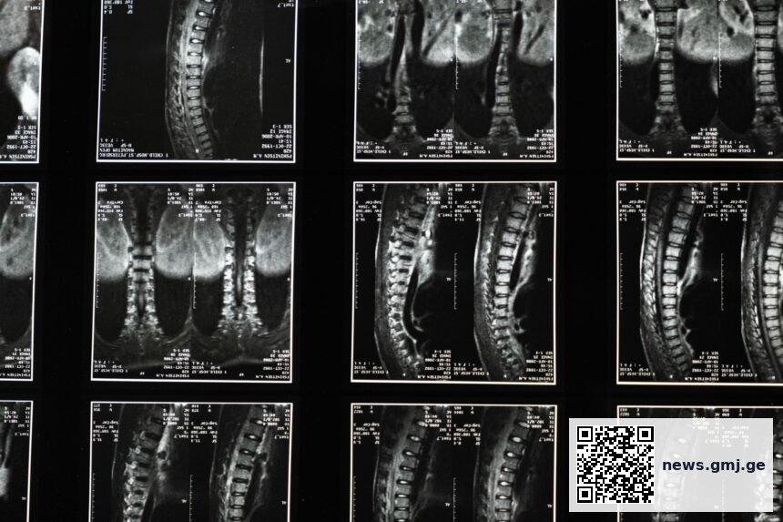
The spine functions as the body’s primary neural communications highway, transmitting signals from the brain to every organ, muscle, and sensory receptor. Rather than merely providing skeletal support, the vertebral column houses the spinal cord and gives rise to 31 pairs of spinal nerves that regulate specific anatomical regions—from respiratory function in the cervical spine to bowel and bladder control via sacral nerves. Understanding this neuroanatomical architecture is essential for clinicians, patients, and public health professionals seeking to address both acute and chronic pain syndromes and systemic dysfunction.
Key takeaways
- The spine houses the spinal cord and gives rise to 31 pairs of segmental nerves, each controlling specific organ systems and body regions
- Compression, irritation, or misalignment of spinal nerves can produce referred pain and dysfunction distant from the site of spinal pathology
- Modifiable risk factors—including posture, core strength, stress, and physical activity—significantly influence spinal nerve function and overall systemic health
- Nerve signal integrity is foundational to optimal nervous system control of movement, respiration, digestion, cardiovascular function, and hormonal regulation
The Spine as Neurological Hub
The human spine consists of 33 vertebrae arranged in five regions: cervical (neck), thoracic (mid-back), lumbar (lower back), sacral (fused base), and coccygeal (tailbone). At each vertebral level, pairs of spinal nerves emerge from the spinal cord through openings called intervertebral foramina, extending to innervate distinct anatomical territories. This segmental organization means that cervical nerves (C1–C8) control the neck, shoulders, and upper extremities; thoracic nerves (T1–T12) innervate the chest wall, organs, and mid-back; lumbar nerves (L1–L5) serve the lower back, hips, and legs; and sacral nerves (S1–S5) regulate pelvic organs, lower limbs, and perineal structures.
When spinal structures—whether vertebrae, discs, ligaments, or muscles—compress or irritate these nerves, the resulting dysfunction extends far beyond local back pain. A compressed cervical nerve root may produce tingling or weakness in the arm; irritation of thoracic nerves can impair digestion or affect breathing; lumbar nerve compression may cause sciatica or lower-limb weakness; and sacral nerve dysfunction can compromise bladder, bowel, and sexual function.

How Spinal Misalignment and Nerve Compression Cascade Through Body Systems
Poor posture, weak core musculature, and sedentary behavior are among the most common culprits in spinal nerve irritation. When the cervical spine is held in sustained forward-flexed posture—a hallmark of modern desk work and smartphone use—the posterior cervical muscles and ligaments bear excessive load, potentially compressing nerve roots and contributing to cervicogenic headaches, neck pain, and upper-extremity paresthesias. Similarly, weak abdominal and gluteal muscles shift load-bearing responsibility to the lumbar spine and its supporting ligaments, increasing disc herniation risk and nerve root irritation.
Psychological stress amplifies these mechanical problems by triggering sustained muscle contraction (myofascial tension) around the spine. This altered muscle tone can further compress neural structures and disrupt the afferent (sensory) signaling that normally modulates pain perception and sympathetic nervous system activity. Prolonged sitting and sedentary behavior reduce blood flow to spinal tissues, promoting inflammation and slowing neural recovery.
Segmental Spinal Nerve Distribution and Common Clinical Presentations
Spinal nerve levels and their primary anatomical territories and organs
Source: Anatomical segmentation based on standard neuroanatomical references | Georgian Medical Journal News
The spinal cord gives rise to 31 pairs of segmental nerves, each innervating specific anatomical regions and organ systems. When a spinal nerve is compressed or irritated, dysfunction and pain can appear in body regions far from the actual site of spinal pathology.
— Standard neuroanatomical principle referenced in clinical anatomy textbooks and neurosurgical literature
Modifiable Risk Factors and Evidence-Based Prevention
Several modifiable lifestyle factors significantly influence spinal health and nerve function. Research published in ergonomics and occupational health journals demonstrates that sustained poor posture increases intradiscal pressure and narrowing of neural foramina. Core strengthening—targeting the deep abdominal stabilizers and gluteal muscles—reduces compensatory stress on the lumbar spine and improves neural signal integrity. Regular physical activity improves spinal tissue blood flow, reduces inflammatory markers, and enhances proprioceptive feedback, all of which support optimal nerve function.
Stress management is increasingly recognized as part of comprehensive spinal health. Chronic psychosocial stress increases sympathetic nervous system tone and muscle guarding, which can exacerbate nerve compression and pain. Conversely, practices that reduce stress—including mindfulness, structured exercise, and adequate sleep—promote parasympathetic tone and allow muscles around the spine to relax, reducing nerve irritation. The World Health Organization identifies stress reduction as a cornerstone of preventive health strategy.
For patients experiencing referred pain—such as sciatic pain originating from lumbar nerve root compression—early recognition of the neurological basis of symptoms is critical. This understanding can redirect care from ineffective local treatments to targeted interventions addressing the actual source of nerve irritation, whether mechanical (posture, weakness, disc herniation) or inflammatory (nerve root edema, ligamentous irritation).
What this means
Frequently asked questions
Can a single misaligned vertebra cause widespread symptoms?
While a single vertebral misalignment does not control your entire body, compression or irritation of the nerve emerging from that level can disrupt function in the specific organs and tissues it innervates. For example, a cervical vertebral misalignment may compress a nerve root, causing arm pain, but not directly affect leg sensation. The key is understanding that nerves follow segmental distribution patterns, so symptoms correlate with spinal level, not with one vertebra controlling all systems.
Why does referred pain occur—pain in one area caused by dysfunction elsewhere on the spine?
Referred pain occurs because the spinal nerve that is irritated sends sensory signals from its entire anatomical territory back to the brain, which may localize the pain sensation to the area normally served by that nerve rather than to the spine itself. For instance, irritation of the L5 nerve root may produce pain felt in the foot or calf, even though the actual problem is at the lumbar spine. This is a normal feature of spinal neuroanatomy and highlights why accurate diagnosis requires clinical evaluation and imaging.
How can I protect my spine and nerve function in daily life?
Maintain neutral spine posture during prolonged sitting; take movement breaks every 30–60 minutes; perform regular core strengthening exercises; engage in consistent aerobic activity to promote spinal tissue blood flow; manage stress through mindfulness or exercise; and ensure adequate sleep. If symptoms develop, early intervention—including physical therapy and ergonomic assessment—can prevent chronic nerve dysfunction. See a clinician if pain, numbness, or weakness persists.
As understanding of spinal neuroanatomy becomes more integrated into public health education and occupational ergonomics standards, both individual and population-level outcomes in pain management and functional mobility are likely to improve. The spine’s role as the body’s primary neural communications system underscores why clinical updates on spinal care and nerve health remain essential for healthcare providers and informed patients alike. Future research should continue to elucidate the long-term effects of modifiable risk factors on spinal nerve function and explore how integrated rehabilitation and workplace interventions can reduce the burden of spinal-related disability globally.
Source: Original educational content on spinal neuroanatomy
🗺️ Find certified Georgian healthcare providers: sheniekimi.ge — Georgia Health Map · 998 clinics · 6,600+ doctors · 2,296 pharmacies
Was this article helpful?
Disclaimer. This article is health journalism intended for general information and education. It is not medical advice and is not a substitute for professional diagnosis or treatment. Always consult a qualified healthcare provider about your individual circumstances. Full disclaimer →
Related Coverage



Editorial standards. This article was produced under the GMJ News editorial process, with oversight by the GMJ Editorial Board. Our editorial process. Spotted an error? Contact the editorial team.